

166
Methods for Setting Up Primary Cultures Specific to Animal Groups
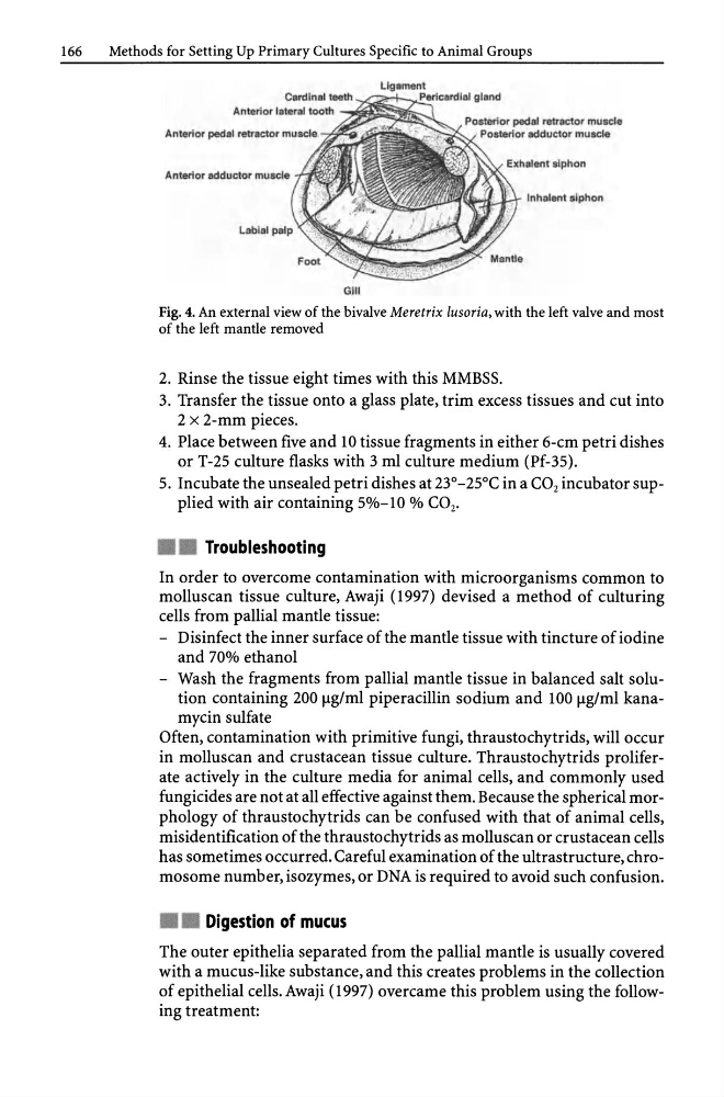
Ligament
Anterior pedal retractor muscle
Anterior adductor muscle fJ~m~k~~~
.,...""'--......- Inhalent siphon
Gill
Fig. 4. An external view of the bivalve Meretrix /usoria, with the left valve and most
of the left mantle removed
2. Rinse the tissue eight times with this MMBSS.
3. Transfer the tissue onto a glass plate, trim excess tissues and cut into
2 x 2-mm pieces.
4. Place between five and 10 tissue fragments in either 6-cm petri dishes
or T-25 culture flasks with 3 ml culture medium (Pf-35).
5. Incubate the unsealed petri dishes at 23°-25°C in a CO 2 incubator supplied with air containing 5%-10 % CO 2 •
• • Troubleshooting
In order to overcome contamination with microorganisms common to
molluscan tissue culture, Awaji (1997) devised a method of culturing
cells from pallial mantle tissue:
- Disinfect the inner surface of the mantle tissue with tincture of iodine
and 70% ethanol
- Wash the fragments from pallial mantle tissue in balanced salt solution containing 200 fig/ml piperacillin sodium and 100 fig/ml kanamycin sulfate
Often, contamination with primitive fungi, thraustochytrids, will occur
in molluscan and crustacean tissue culture. Thraustochytrids proliferate actively in the culture media for animal cells, and commonly used
fungicides are not at all effective against them. Because the spherical morphology of thraustochytrids can be confused with that of animal cells,
misidentification of the thraustochytrids as molluscan or crustacean cells
has sometimes occurred. Careful examination of the ultrastructure, chromosome number, isozymes, or DNA is required to avoid such confusion .
• • Digestion of mucus
The outer epithelia separated from the pallial mantle is usually covered
with a mucus-like substance, and this creates problems in the collection
of epithelial cells. Awaji (1997) overcame this problem using the following treatment:
Methods for Setting Up Primary Cultures Specific to Animal Groups
Ligament
Anterior pedal retractor muscle
Anterior adductor muscle fJ~m~k~~~
.,...""'--......- Inhalent siphon
Gill
Fig. 4. An external view of the bivalve Meretrix /usoria, with the left valve and most
of the left mantle removed
2. Rinse the tissue eight times with this MMBSS.
3. Transfer the tissue onto a glass plate, trim excess tissues and cut into
2 x 2-mm pieces.
4. Place between five and 10 tissue fragments in either 6-cm petri dishes
or T-25 culture flasks with 3 ml culture medium (Pf-35).
5. Incubate the unsealed petri dishes at 23°-25°C in a CO 2 incubator supplied with air containing 5%-10 % CO 2 •
• • Troubleshooting
In order to overcome contamination with microorganisms common to
molluscan tissue culture, Awaji (1997) devised a method of culturing
cells from pallial mantle tissue:
- Disinfect the inner surface of the mantle tissue with tincture of iodine
and 70% ethanol
- Wash the fragments from pallial mantle tissue in balanced salt solution containing 200 fig/ml piperacillin sodium and 100 fig/ml kanamycin sulfate
Often, contamination with primitive fungi, thraustochytrids, will occur
in molluscan and crustacean tissue culture. Thraustochytrids proliferate actively in the culture media for animal cells, and commonly used
fungicides are not at all effective against them. Because the spherical morphology of thraustochytrids can be confused with that of animal cells,
misidentification of the thraustochytrids as molluscan or crustacean cells
has sometimes occurred. Careful examination of the ultrastructure, chromosome number, isozymes, or DNA is required to avoid such confusion .
• • Digestion of mucus
The outer epithelia separated from the pallial mantle is usually covered
with a mucus-like substance, and this creates problems in the collection
of epithelial cells. Awaji (1997) overcame this problem using the following treatment: