

14 Echinodermata
1SS
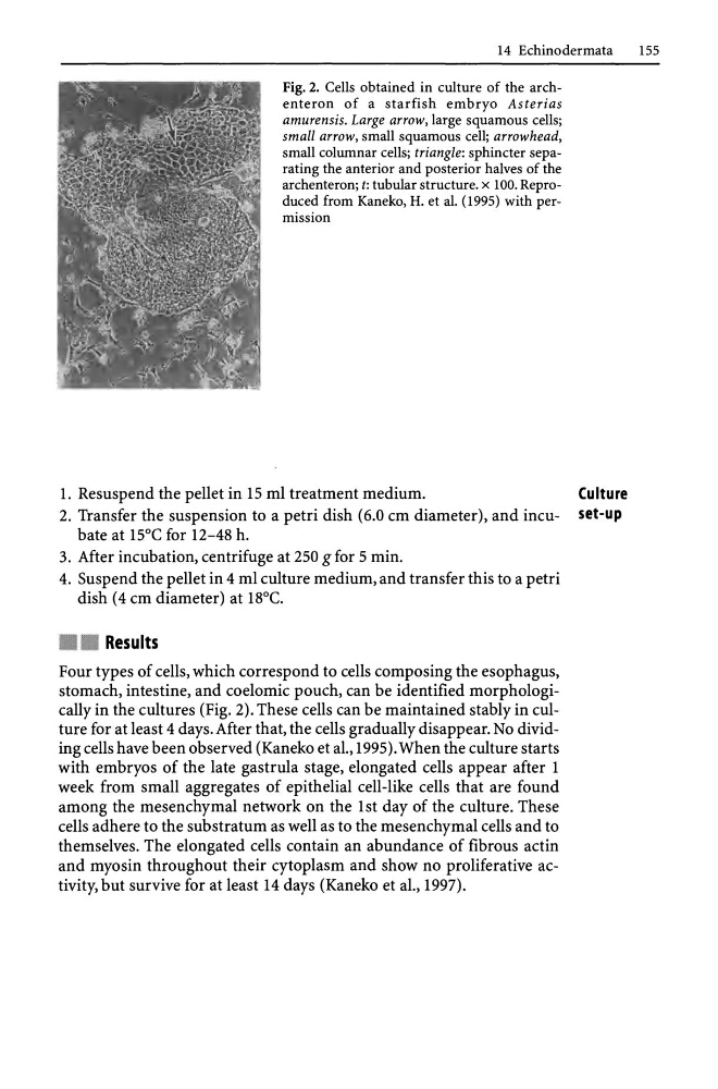
Fig. 2. Cells obtained in culture of the archenteron of a starfish embryo Asterias
amurensis. Large arrow, large squamous cells;
small arrow, small squamous cell; arrowhead,
small columnar cells; triangle: sphincter separating the anterior and posterior halves of the
archenteron; t: tubular structure. x 100. Reproduced from Kaneko, H. et al. (199S) with permission
1. Resuspend the pellet in 15 ml treatment medium.
Culture
2. Transfer the suspension to a petri dish (6.0 cm diameter), and incu- set-up
bate at 15°C for 12-48 h.
3. After incubation, centrifuge at 250 g for 5 min.
4. Suspend the pellet in 4 ml culture medium, and transfer this to a petri
dish (4 cm diameter) at 18°C .
• Results
Four types of cells, which correspond to cells composing the esophagus,
stomach, intestine, and coelomic pouch, can be identified morphologically in the cultures (Fig. 2). These cells can be maintained stably in culture for at least 4 days. After that, the cells gradually disappear. No dividing cells have been observed (Kaneko et aI., 1995). When the culture starts
with embryos of the late gastrula stage, elongated cells appear after 1
week from small aggregates of epithelial cell-like cells that are found
among the mesenchymal network on the 1st day of the culture. These
cells adhere to the substratum as well as to the mesenchymal cells and to
themselves. The elongated cells contain an abundance of fibrous actin
and myosin throughout their cytoplasm and show no proliferative activity, but survive for at least 14 days (Kaneko et aI., 1997).
1SS
Fig. 2. Cells obtained in culture of the archenteron of a starfish embryo Asterias
amurensis. Large arrow, large squamous cells;
small arrow, small squamous cell; arrowhead,
small columnar cells; triangle: sphincter separating the anterior and posterior halves of the
archenteron; t: tubular structure. x 100. Reproduced from Kaneko, H. et al. (199S) with permission
1. Resuspend the pellet in 15 ml treatment medium.
Culture
2. Transfer the suspension to a petri dish (6.0 cm diameter), and incu- set-up
bate at 15°C for 12-48 h.
3. After incubation, centrifuge at 250 g for 5 min.
4. Suspend the pellet in 4 ml culture medium, and transfer this to a petri
dish (4 cm diameter) at 18°C .
• Results
Four types of cells, which correspond to cells composing the esophagus,
stomach, intestine, and coelomic pouch, can be identified morphologically in the cultures (Fig. 2). These cells can be maintained stably in culture for at least 4 days. After that, the cells gradually disappear. No dividing cells have been observed (Kaneko et aI., 1995). When the culture starts
with embryos of the late gastrula stage, elongated cells appear after 1
week from small aggregates of epithelial cell-like cells that are found
among the mesenchymal network on the 1st day of the culture. These
cells adhere to the substratum as well as to the mesenchymal cells and to
themselves. The elongated cells contain an abundance of fibrous actin
and myosin throughout their cytoplasm and show no proliferative activity, but survive for at least 14 days (Kaneko et aI., 1997).