

13 Prochordata
147
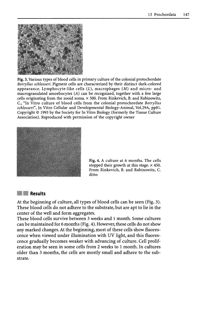
Fig. 3. Various types of blood cells in primary culture of the colonial protochordate
Botryllus schlosseri. Pigment cells are characterized by their distinct dark-colored
appearance. Lymphocyte-like cells (L), macrophages (M) and micro- and
macro granulated amoebocytes (A) can be recognized, together with a few large
cells originating from the zooid soma. x 500. From Rinkevich, B. and Rabinowitz,
c., "In Vitro culture of blood cells from the colonial protochordate Botryllus
schlosseri", In Vitro Cellular and Developmental Biology-Animal, Vo1.29A, pp81.
Copyright © 1993 by the Society for In Vitro Biology (formerly the Tissue Culture
Association). Reproduced with permission of the copyright owner
• • Results
Fig. 4. A culture at 6 months. The cells
stopped their growth at this stage. x 450.
From Rinkevich, B. and Rabinowitz, C.
ditto
At the beginning of culture, all types of blood cells can be seen (Fig. 3).
These blood cells do not adhere to the substrate, but are apt to lie in the
center of the well and form aggregates.
These blood cells survive between 3 weeks and 1 month. Some cultures
can be maintained for 6 months (Fig. 4). However, these cells do not show
any marked changes. At the beginning, most of these cells show fluorescence when viewed under illumination with UV light, and this fluorescence gradually becomes weaker with advancing of culture. Cell proliferation may be seen in some cells from 2 weeks to 1 month. In cultures
older than 3 months, the cells are mostly small and adhere to the substrate.
147
Fig. 3. Various types of blood cells in primary culture of the colonial protochordate
Botryllus schlosseri. Pigment cells are characterized by their distinct dark-colored
appearance. Lymphocyte-like cells (L), macrophages (M) and micro- and
macro granulated amoebocytes (A) can be recognized, together with a few large
cells originating from the zooid soma. x 500. From Rinkevich, B. and Rabinowitz,
c., "In Vitro culture of blood cells from the colonial protochordate Botryllus
schlosseri", In Vitro Cellular and Developmental Biology-Animal, Vo1.29A, pp81.
Copyright © 1993 by the Society for In Vitro Biology (formerly the Tissue Culture
Association). Reproduced with permission of the copyright owner
• • Results
Fig. 4. A culture at 6 months. The cells
stopped their growth at this stage. x 450.
From Rinkevich, B. and Rabinowitz, C.
ditto
At the beginning of culture, all types of blood cells can be seen (Fig. 3).
These blood cells do not adhere to the substrate, but are apt to lie in the
center of the well and form aggregates.
These blood cells survive between 3 weeks and 1 month. Some cultures
can be maintained for 6 months (Fig. 4). However, these cells do not show
any marked changes. At the beginning, most of these cells show fluorescence when viewed under illumination with UV light, and this fluorescence gradually becomes weaker with advancing of culture. Cell proliferation may be seen in some cells from 2 weeks to 1 month. In cultures
older than 3 months, the cells are mostly small and adhere to the substrate.