

12 Arthropods Other than Insects
129
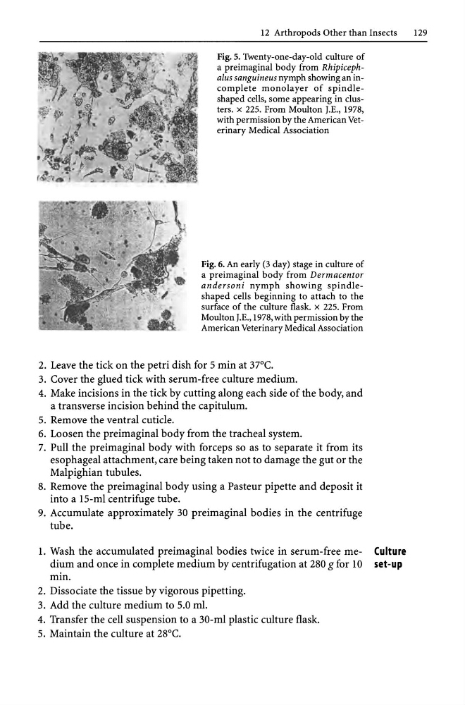
Fig. 5. Twenty-one-day-old culture of
a preimaginal body from Rhipicephalus sanguineus nymph showing an incomplete monolayer of spindleshaped cells, some appearing in clusters. x 225. From Moulton J.E., 1978,
with permission by the American Veterinary Medical Association
Fig. 6. An early (3 day) stage in culture of
a preimaginal body from Dermacentor
andersoni nymph showing spindleshaped cells beginning to attach to the
surface of the culture flask. x 225. From
Moulton J.E., 1978, with permission by the
American Veterinary Medical Association
2. Leave the tick on the petri dish for 5 min at 37°C.
3. Cover the glued tick with serum-free culture medium.
4. Make incisions in the tick by cutting along each side of the body, and
a transverse incision behind the capitulum.
5. Remove the ventral cuticle.
6. Loosen the pre imaginal body from the tracheal system.
7. Pull the pre imaginal body with forceps so as to separate it from its
esophageal attachment, care being taken not to damage the gut or the
Malpighian tubules.
8. Remove the preimaginal body using a Pasteur pipette and deposit it
into a IS-ml centrifuge tube.
9. Accumulate approximately 30 preimaginal bodies in the centrifuge
tube.
1. Wash the accumulated preimaginal bodies twice in serum-free me- Culture
dium and once in complete medium by centrifugation at 280 g for 10 set-up
min.
2. Dissociate the tissue by vigorous pipetting.
3. Add the culture medium to 5.0 ml.
4. Transfer the cell suspension to a 30-ml plastic culture flask.
5. Maintain the culture at 28°C.
129
Fig. 5. Twenty-one-day-old culture of
a preimaginal body from Rhipicephalus sanguineus nymph showing an incomplete monolayer of spindleshaped cells, some appearing in clusters. x 225. From Moulton J.E., 1978,
with permission by the American Veterinary Medical Association
Fig. 6. An early (3 day) stage in culture of
a preimaginal body from Dermacentor
andersoni nymph showing spindleshaped cells beginning to attach to the
surface of the culture flask. x 225. From
Moulton J.E., 1978, with permission by the
American Veterinary Medical Association
2. Leave the tick on the petri dish for 5 min at 37°C.
3. Cover the glued tick with serum-free culture medium.
4. Make incisions in the tick by cutting along each side of the body, and
a transverse incision behind the capitulum.
5. Remove the ventral cuticle.
6. Loosen the pre imaginal body from the tracheal system.
7. Pull the pre imaginal body with forceps so as to separate it from its
esophageal attachment, care being taken not to damage the gut or the
Malpighian tubules.
8. Remove the preimaginal body using a Pasteur pipette and deposit it
into a IS-ml centrifuge tube.
9. Accumulate approximately 30 preimaginal bodies in the centrifuge
tube.
1. Wash the accumulated preimaginal bodies twice in serum-free me- Culture
dium and once in complete medium by centrifugation at 280 g for 10 set-up
min.
2. Dissociate the tissue by vigorous pipetting.
3. Add the culture medium to 5.0 ml.
4. Transfer the cell suspension to a 30-ml plastic culture flask.
5. Maintain the culture at 28°C.