

124
Methods for Setting Up Primary Cultures Specific to Animal Groups
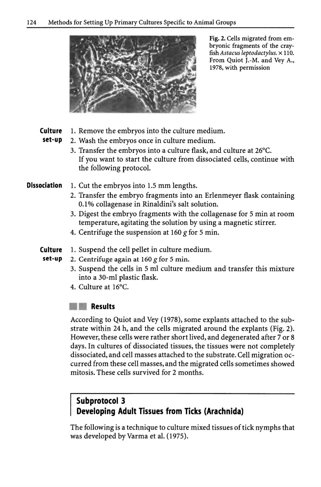
Fig. 2. Cells migrated from embryonic fragments of the crayfish Astacus leptodactylus. x llO.
From Quiot J.-M. and Vey A.,
1978, with permission
Culture 1. Remove the embryos into the culture medium.
set-up 2. Wash the embryos once in culture medium.
3. Transfer the embryos into a culture flask, and culture at 26°C.
If you want to start the culture from dissociated cells, continue with
the following protocol.
Dissociation 1. Cut the embryos into 1.5 mm lengths.
2. Transfer the embryo fragments into an Erlenmeyer flask containing
0.1 % collagenase in Rinaldini's salt solution.
3. Digest the embryo fragments with the collagenase for 5 min at room
temperature, agitating the solution by using a magnetic stirrer.
4. Centrifuge the suspension at 160 g for 5 min.
Culture 1. Suspend the cell pellet in culture medium.
set-up 2. Centrifuge again at 160 g for 5 min.
3. Suspend the cells in 5 ml culture medium and transfer this mixture
into a 30-ml plastic flask.
4. Culture at 16°C.
• • Results
According to Quiot and Vey (1978), some explants attached to the substrate within 24 h, and the cells migrated around the explants (Fig. 2).
However, these cells were rather short lived, and degenerated after 7 or 8
days. In cultures of dissociated tissues, the tissues were not completely
dissociated, and cell masses attached to the substrate. Cell migration occurred from these cell masses, and the migrated cells sometimes showed
mitosis. These cells survived for 2 months.
Subprotocol 3
Developing Adult Tissues from Ticks (Arachnida)
The following is a technique to culture mixed tissues of tick nymphs that
was developed by Varma et al. (1975).
Methods for Setting Up Primary Cultures Specific to Animal Groups
Fig. 2. Cells migrated from embryonic fragments of the crayfish Astacus leptodactylus. x llO.
From Quiot J.-M. and Vey A.,
1978, with permission
Culture 1. Remove the embryos into the culture medium.
set-up 2. Wash the embryos once in culture medium.
3. Transfer the embryos into a culture flask, and culture at 26°C.
If you want to start the culture from dissociated cells, continue with
the following protocol.
Dissociation 1. Cut the embryos into 1.5 mm lengths.
2. Transfer the embryo fragments into an Erlenmeyer flask containing
0.1 % collagenase in Rinaldini's salt solution.
3. Digest the embryo fragments with the collagenase for 5 min at room
temperature, agitating the solution by using a magnetic stirrer.
4. Centrifuge the suspension at 160 g for 5 min.
Culture 1. Suspend the cell pellet in culture medium.
set-up 2. Centrifuge again at 160 g for 5 min.
3. Suspend the cells in 5 ml culture medium and transfer this mixture
into a 30-ml plastic flask.
4. Culture at 16°C.
• • Results
According to Quiot and Vey (1978), some explants attached to the substrate within 24 h, and the cells migrated around the explants (Fig. 2).
However, these cells were rather short lived, and degenerated after 7 or 8
days. In cultures of dissociated tissues, the tissues were not completely
dissociated, and cell masses attached to the substrate. Cell migration occurred from these cell masses, and the migrated cells sometimes showed
mitosis. These cells survived for 2 months.
Subprotocol 3
Developing Adult Tissues from Ticks (Arachnida)
The following is a technique to culture mixed tissues of tick nymphs that
was developed by Varma et al. (1975).