

110
Methods for Setting Up Primary Cultures Specific to Animal Groups
Sterilization
Dissection of
embryonic
!rvous organs
Dissociation
Culture
set-up
Dissection of
embryonic
foregut
MedJum
DiMociated DCrve oelb
Forecut esplant
II. Procedure
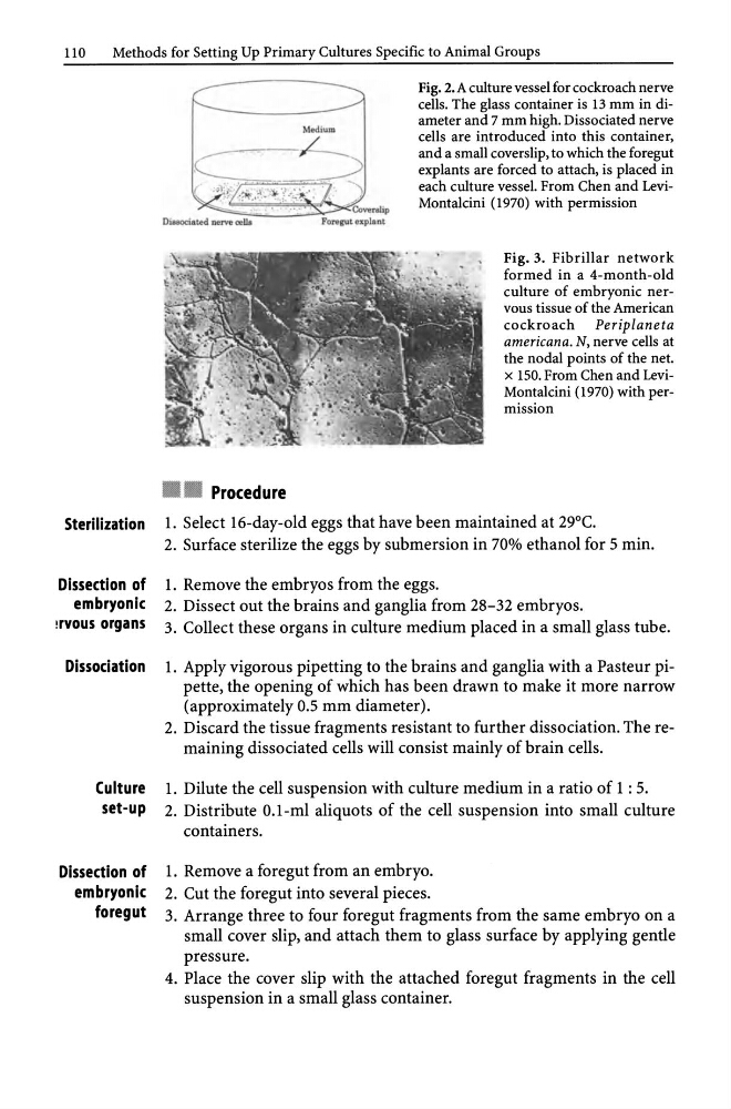
Fig. 2. A culture vessel for cockroach nerve
cells. The glass container is 13 mm in diameter and 7 mm high. Dissociated nerve
cells are introduced into this container,
and a small coverslip, to which the foregut
explants are forced to attach, is placed in
each culture vessel. From Chen and LeviMontalcini (1970) with permission
Fig. 3. Fibrillar network
formed in a 4-month-old
culture of embryonic nervous tissue of the American
cockroach Periplaneta
americana. N, nerve cells at
the nodal points of the net.
x 150. From Chen and LeviMontalcini (1970) with permission
l. Select 16-day-old eggs that have been maintained at 29°C.
2. Surface sterilize the eggs by submersion in 70% ethanol for 5 min.
1. Remove the embryos from the eggs.
2. Dissect out the brains and ganglia from 28-32 embryos.
3. Collect these organs in culture medium placed in a small glass tube.
l. Apply vigorous pipetting to the brains and ganglia with a Pasteur pipette, the opening of which has been drawn to make it more narrow
(approximately 0.5 mm diameter).
2. Discard the tissue fragments resistant to further dissociation. The remaining dissociated cells will consist mainly of brain cells.
l. Dilute the cell suspension with culture medium in a ratio of 1 : 5.
2. Distribute O.I-ml aliquots of the cell suspension into small culture
containers.
1. Remove a foregut from an embryo.
2. Cut the foregut into several pieces.
3. Arrange three to four foregut fragments from the same embryo on a
small cover slip, and attach them to glass surface by applying gentle
pressure.
4. Place the cover slip with the attached foregut fragments in the cell
suspension in a small glass container.
Methods for Setting Up Primary Cultures Specific to Animal Groups
Sterilization
Dissection of
embryonic
!rvous organs
Dissociation
Culture
set-up
Dissection of
embryonic
foregut
MedJum
DiMociated DCrve oelb
Forecut esplant
II. Procedure
Fig. 2. A culture vessel for cockroach nerve
cells. The glass container is 13 mm in diameter and 7 mm high. Dissociated nerve
cells are introduced into this container,
and a small coverslip, to which the foregut
explants are forced to attach, is placed in
each culture vessel. From Chen and LeviMontalcini (1970) with permission
Fig. 3. Fibrillar network
formed in a 4-month-old
culture of embryonic nervous tissue of the American
cockroach Periplaneta
americana. N, nerve cells at
the nodal points of the net.
x 150. From Chen and LeviMontalcini (1970) with permission
l. Select 16-day-old eggs that have been maintained at 29°C.
2. Surface sterilize the eggs by submersion in 70% ethanol for 5 min.
1. Remove the embryos from the eggs.
2. Dissect out the brains and ganglia from 28-32 embryos.
3. Collect these organs in culture medium placed in a small glass tube.
l. Apply vigorous pipetting to the brains and ganglia with a Pasteur pipette, the opening of which has been drawn to make it more narrow
(approximately 0.5 mm diameter).
2. Discard the tissue fragments resistant to further dissociation. The remaining dissociated cells will consist mainly of brain cells.
l. Dilute the cell suspension with culture medium in a ratio of 1 : 5.
2. Distribute O.I-ml aliquots of the cell suspension into small culture
containers.
1. Remove a foregut from an embryo.
2. Cut the foregut into several pieces.
3. Arrange three to four foregut fragments from the same embryo on a
small cover slip, and attach them to glass surface by applying gentle
pressure.
4. Place the cover slip with the attached foregut fragments in the cell
suspension in a small glass container.