

15
gonad develops on the basal zooidal wall, usually spreading to
adjoining areas of the lateral and transverse walls. More rarely
it occupies most or all of the surface of the basal wall (except
the area occupied by the ovary in the case of hermaphrodite
zooids). In several species, spermatogenic tissue lies, completely or partly, on the inner surface of the frontal wall/shield.
In “Calyptotheca” variolosa , developing male gametes were
found on the surface of the lower wall of the compensation
sac. In several species they were noted both on the zooidal
wall and on funicular cords. In three species, spermatogenic
tissue lines the entire proximal part of the cystid, being located
on the cystid wall and the adjoining parts of funicular cords.
A similar variant was found in Calyptotheca triangula but in
this species spermatogenic tissue lined not only the proximal
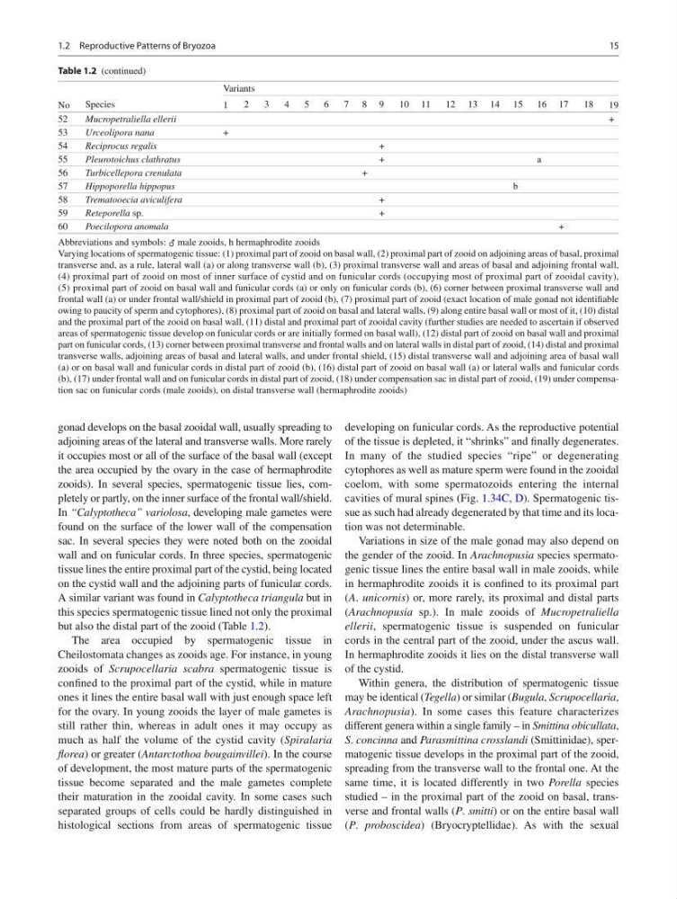
but also the distal part of the zooid (Table 1.2 ).
The area occupied by spermatogenic tissue in
Cheilostomata changes as zooids age. For instance, in young
zooids of Scrupocellaria scabra spermatogenic tissue is
confi ned to the proximal part of the cystid, while in mature
ones it lines the entire basal wall with just enough space left
for the ovary. In young zooids the layer of male gametes is
still rather thin, whereas in adult ones it may occupy as
much as half the volume of the cystid cavity ( Spiralaria
fl orea ) or greater ( Antarctothoa bougainvillei ). In the course
of development, the most mature parts of the spermatogenic
tissue become separated and the male gametes complete
their maturation in the zooidal cavity. In some cases such
separated groups of cells could be hardly distinguished in
histological sections from areas of spermatogenic tissue
developing on funicular cords. As the reproductive potential
of the tissue is depleted, it “shrinks” and fi nally degenerates.
In many of the studied species “ripe” or degenerating
cytophores as well as mature sperm were found in the zooidal
coelom, with some spermatozoids entering the internal
cavities of mural spines (Fig. 1.34C, D ). Spermatogenic tissue as such had already degenerated by that time and its location was not determinable.
Variations in size of the male gonad may also depend on
the gender of the zooid. In Arachnopusia species spermatogenic tissue lines the entire basal wall in male zooids, while
in hermaphrodite zooids it is confi ned to its proximal part
( A . unicornis ) or, more rarely, its proximal and distal parts
( Arachnopusia sp.). In male zooids of Mucropetraliella
ellerii , spermatogenic tissue is suspended on funicular
cords in the central part of the zooid, under the ascus wall.
In hermaphrodite zooids it lies on the distal transverse wall
of the cystid.
Within genera, the distribution of spermatogenic tissue
may be identical ( Tegella ) or similar ( Bugula , Scrupocellaria ,
Arachnopusia ). In some cases this feature characterizes
different genera within a single family – in Smittina obicullata ,
S . concinna and Parasmittina crosslandi (Smittinidae), spermatogenic tissue develops in the proximal part of the zooid,
spreading from the transverse wall to the frontal one. At the
same time, it is located differently in two Porella species
studied – in the proximal part of the zooid on basal, transverse and frontal walls ( P . smitti ) or on the entire basal wall
( P . proboscidea ) (Bryocryptellidae). As with the sexual
No
Species
Variants
1
2
3
4
5
6
7 8 9
10 11
12 13 14 15 16 17
18
19
52
Mucropetraliella ellerii
+
53
Urceolipora nana
+
54
Reciprocus regalis
+
55
Pleurotoichus clathratus
+
a
56
Turbicellepora crenulata
+
57
Hippoporella hippopus
b
58
Trematooecia aviculifera
+
59
Reteporella sp.
+
60
Poecilopora anomala
+
Abbreviations and symbols: ♂ male zooids, h hermaphrodite zooids
Varying locations of spermatogenic tissue: (1) proximal part of zooid on basal wall, (2) proximal part of zooid on adjoining areas of basal, proximal
transverse and, as a rule, lateral wall (a) or along transverse wall (b), (3) proximal transverse wall and areas of basal and adjoining frontal wall,
(4) proximal part of zooid on most of inner surface of cystid and on funicular cords (occupying most of proximal part of zooidal cavity),
(5) proximal part of zooid on basal wall and funicular cords (a) or only on funicular cords (b), (6) corner between proximal transverse wall and
frontal wall (a) or under frontal wall/shield in proximal part of zooid (b), (7) proximal part of zooid (exact location of male gonad not identifi able
owing to paucity of sperm and cytophores), (8) proximal part of zooid on basal and lateral walls, (9) along entire basal wall or most of it, (10) distal
and the proximal part of the zooid on basal wall, (11) distal and proximal part of zooidal cavity (further studies are needed to ascertain if observed
areas of spermatogenic tissue develop on funicular cords or are initially formed on basal wall), (12) distal part of zooid on basal wall and proximal
part on funicular cords, (13) corner between proximal transverse and frontal walls and on lateral walls in distal part of zooid, (14) distal and proximal
transverse walls, adjoining areas of basal and lateral walls, and under frontal shield, (15) distal transverse wall and adjoining area of basal wall
(a) or on basal wall and funicular cords in distal part of zooid (b), (16) distal part of zooid on basal wall (a) or lateral walls and funicular cords
(b), (17) under frontal wall and on funicular cords in distal part of zooid, (18) under compensation sac in distal part of zooid, (19) under compensation sac on funicular cords (male zooids), on distal transverse wall (hermaphrodite zooids)
Table 1.2 (continued)
1.2 Reproductive Patterns of Bryozoa
gonad develops on the basal zooidal wall, usually spreading to
adjoining areas of the lateral and transverse walls. More rarely
it occupies most or all of the surface of the basal wall (except
the area occupied by the ovary in the case of hermaphrodite
zooids). In several species, spermatogenic tissue lies, completely or partly, on the inner surface of the frontal wall/shield.
In “Calyptotheca” variolosa , developing male gametes were
found on the surface of the lower wall of the compensation
sac. In several species they were noted both on the zooidal
wall and on funicular cords. In three species, spermatogenic
tissue lines the entire proximal part of the cystid, being located
on the cystid wall and the adjoining parts of funicular cords.
A similar variant was found in Calyptotheca triangula but in
this species spermatogenic tissue lined not only the proximal
but also the distal part of the zooid (Table 1.2 ).
The area occupied by spermatogenic tissue in
Cheilostomata changes as zooids age. For instance, in young
zooids of Scrupocellaria scabra spermatogenic tissue is
confi ned to the proximal part of the cystid, while in mature
ones it lines the entire basal wall with just enough space left
for the ovary. In young zooids the layer of male gametes is
still rather thin, whereas in adult ones it may occupy as
much as half the volume of the cystid cavity ( Spiralaria
fl orea ) or greater ( Antarctothoa bougainvillei ). In the course
of development, the most mature parts of the spermatogenic
tissue become separated and the male gametes complete
their maturation in the zooidal cavity. In some cases such
separated groups of cells could be hardly distinguished in
histological sections from areas of spermatogenic tissue
developing on funicular cords. As the reproductive potential
of the tissue is depleted, it “shrinks” and fi nally degenerates.
In many of the studied species “ripe” or degenerating
cytophores as well as mature sperm were found in the zooidal
coelom, with some spermatozoids entering the internal
cavities of mural spines (Fig. 1.34C, D ). Spermatogenic tissue as such had already degenerated by that time and its location was not determinable.
Variations in size of the male gonad may also depend on
the gender of the zooid. In Arachnopusia species spermatogenic tissue lines the entire basal wall in male zooids, while
in hermaphrodite zooids it is confi ned to its proximal part
( A . unicornis ) or, more rarely, its proximal and distal parts
( Arachnopusia sp.). In male zooids of Mucropetraliella
ellerii , spermatogenic tissue is suspended on funicular
cords in the central part of the zooid, under the ascus wall.
In hermaphrodite zooids it lies on the distal transverse wall
of the cystid.
Within genera, the distribution of spermatogenic tissue
may be identical ( Tegella ) or similar ( Bugula , Scrupocellaria ,
Arachnopusia ). In some cases this feature characterizes
different genera within a single family – in Smittina obicullata ,
S . concinna and Parasmittina crosslandi (Smittinidae), spermatogenic tissue develops in the proximal part of the zooid,
spreading from the transverse wall to the frontal one. At the
same time, it is located differently in two Porella species
studied – in the proximal part of the zooid on basal, transverse and frontal walls ( P . smitti ) or on the entire basal wall
( P . proboscidea ) (Bryocryptellidae). As with the sexual
No
Species
Variants
1
2
3
4
5
6
7 8 9
10 11
12 13 14 15 16 17
18
19
52
Mucropetraliella ellerii
+
53
Urceolipora nana
+
54
Reciprocus regalis
+
55
Pleurotoichus clathratus
+
a
56
Turbicellepora crenulata
+
57
Hippoporella hippopus
b
58
Trematooecia aviculifera
+
59
Reteporella sp.
+
60
Poecilopora anomala
+
Abbreviations and symbols: ♂ male zooids, h hermaphrodite zooids
Varying locations of spermatogenic tissue: (1) proximal part of zooid on basal wall, (2) proximal part of zooid on adjoining areas of basal, proximal
transverse and, as a rule, lateral wall (a) or along transverse wall (b), (3) proximal transverse wall and areas of basal and adjoining frontal wall,
(4) proximal part of zooid on most of inner surface of cystid and on funicular cords (occupying most of proximal part of zooidal cavity),
(5) proximal part of zooid on basal wall and funicular cords (a) or only on funicular cords (b), (6) corner between proximal transverse wall and
frontal wall (a) or under frontal wall/shield in proximal part of zooid (b), (7) proximal part of zooid (exact location of male gonad not identifi able
owing to paucity of sperm and cytophores), (8) proximal part of zooid on basal and lateral walls, (9) along entire basal wall or most of it, (10) distal
and the proximal part of the zooid on basal wall, (11) distal and proximal part of zooidal cavity (further studies are needed to ascertain if observed
areas of spermatogenic tissue develop on funicular cords or are initially formed on basal wall), (12) distal part of zooid on basal wall and proximal
part on funicular cords, (13) corner between proximal transverse and frontal walls and on lateral walls in distal part of zooid, (14) distal and proximal
transverse walls, adjoining areas of basal and lateral walls, and under frontal shield, (15) distal transverse wall and adjoining area of basal wall
(a) or on basal wall and funicular cords in distal part of zooid (b), (16) distal part of zooid on basal wall (a) or lateral walls and funicular cords
(b), (17) under frontal wall and on funicular cords in distal part of zooid, (18) under compensation sac in distal part of zooid, (19) under compensation sac on funicular cords (male zooids), on distal transverse wall (hermaphrodite zooids)
Table 1.2 (continued)
1.2 Reproductive Patterns of Bryozoa