

234
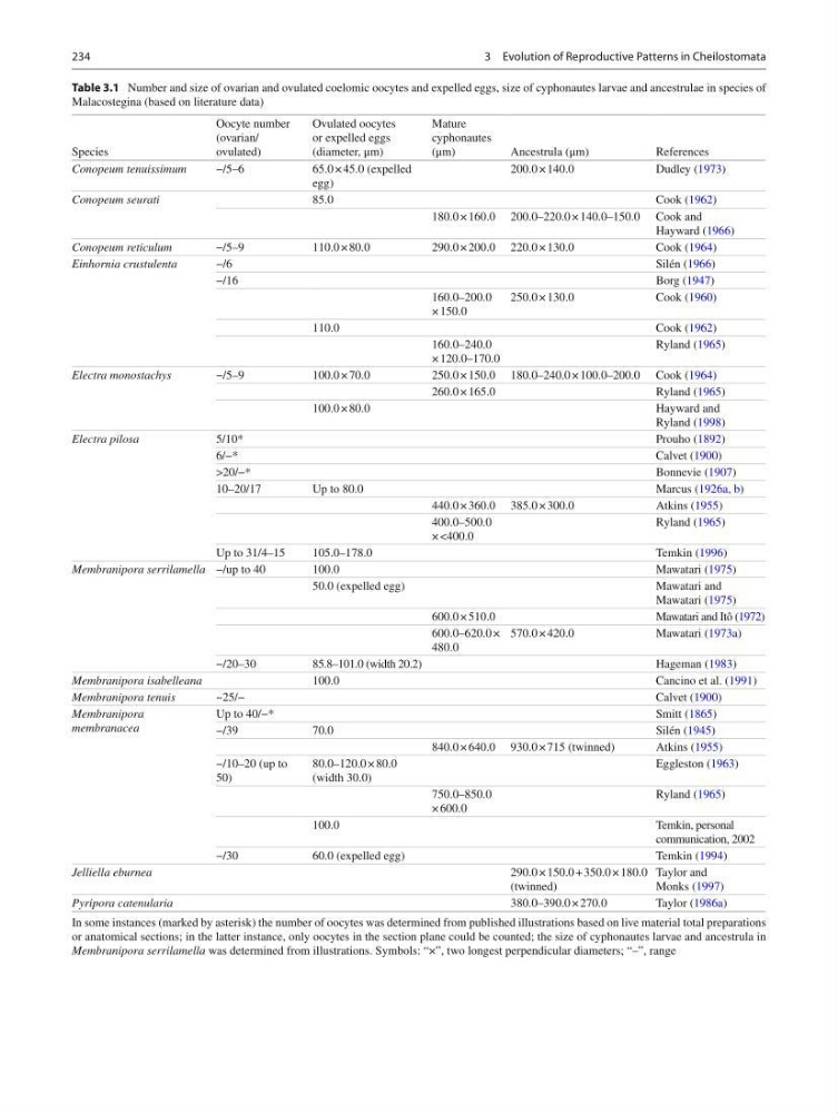
Table 3.1 Number and size of ovarian and ovulated coelomic oocytes and expelled eggs, size of cyphonautes larvae and ancestrulae in species of
Malacostegina (based on literature data)
Species
Oocyte number
(ovarian/
ovulated)
Ovulated oocytes
or expelled eggs
(diameter, μm)
Mature
cyphonautes
(μm)
Ancestrula (μm)
References
Conopeum tenuissimum
−/5–6
65.0 × 45.0 (expelled
egg)
200.0 × 140.0
Dudley ( 1973 )
Conopeum seurati
85.0
Cook ( 1962 )
180.0 × 160.0 200.0–220.0 × 140.0–150.0 Cook and
Hayward ( 1966 )
Conopeum reticulum
−/5–9
110.0 × 80.0
290.0 × 200.0 220.0 × 130.0
Cook ( 1964 )
Einhornia crustulenta
−/6
Silén ( 1966 )
−/16
Borg ( 1947 )
160.0–200.0
× 150.0
250.0 × 130.0
Cook ( 1960 )
110.0
Cook ( 1962 )
160.0–240.0
× 120.0–170.0
Ryland ( 1965 )
Electra monostachys
−/5–9
100.0 × 70.0
250.0 × 150.0 180.0–240.0 × 100.0–200.0 Cook ( 1964 )
260.0 × 165.0
Ryland ( 1965 )
100.0 × 80.0
Hayward and
Ryland ( 1998 )
Electra pilosa
5/10*
Prouho ( 1892 )
6/−*
Calvet ( 1900 )
>20/−*
Bonnevie ( 1907 )
10–20/17
Up to 80.0
Marcus ( 1926a , b )
440.0 × 360.0 385.0 × 300.0
Atkins ( 1955 )
400.0–500.0
× <400.0
Ryland ( 1965 )
Up to 31/4–15
105.0–178.0
Temkin ( 1996 )
Membranipora serrilamella −/up to 40
100.0
Mawatari ( 1975 )
50.0 (expelled egg)
Mawatari and
Mawatari ( 1975 )
600.0 × 510.0
Mawatari and Itô ( 1972 )
600.0–620.0 ×
480.0
570.0 × 420.0
Mawatari ( 1973a )
−/20–30
85.8–101.0 (width 20.2)
Hageman ( 1983 )
Membranipora isabelleana
100.0
Cancino et al. ( 1991 )
Membranipora tenuis
~25/−
Calvet ( 1900 )
Membranipora
membranacea
Up to 40/−*
Smitt ( 1865 )
−/39
70.0
Silén ( 1945 )
840.0 × 640.0 930.0 × 715 (twinned)
Atkins ( 1955 )
−/10–20 (up to
50)
80.0–120.0 × 80.0
(width 30.0)
Eggleston ( 1963 )
750.0–850.0
× 600.0
Ryland ( 1965 )
100.0
Temkin, personal
communication, 2002
−/30
60.0 (expelled egg)
Temkin ( 1994 )
Jelliella eburnea
290.0 × 150.0 + 350.0 × 180.0
(twinned)
Taylor and
Monks ( 1997 )
Pyripora catenularia
380.0–390.0 × 270.0
Taylor ( 1986a )
In some instances (marked by asterisk) the number of oocytes was determined from published illustrations based on live material total preparations
or anatomical sections; in the latter instance, only oocytes in the section plane could be counted; the size of cyphonautes larvae and ancestrula in
Membranipora serrilamella was determined from illustrations. Symbols: “×”, two longest perpendicular diameters; “–”, range
3 Evolution of Reproductive Patterns in Cheilostomata
Table 3.1 Number and size of ovarian and ovulated coelomic oocytes and expelled eggs, size of cyphonautes larvae and ancestrulae in species of
Malacostegina (based on literature data)
Species
Oocyte number
(ovarian/
ovulated)
Ovulated oocytes
or expelled eggs
(diameter, μm)
Mature
cyphonautes
(μm)
Ancestrula (μm)
References
Conopeum tenuissimum
−/5–6
65.0 × 45.0 (expelled
egg)
200.0 × 140.0
Dudley ( 1973 )
Conopeum seurati
85.0
Cook ( 1962 )
180.0 × 160.0 200.0–220.0 × 140.0–150.0 Cook and
Hayward ( 1966 )
Conopeum reticulum
−/5–9
110.0 × 80.0
290.0 × 200.0 220.0 × 130.0
Cook ( 1964 )
Einhornia crustulenta
−/6
Silén ( 1966 )
−/16
Borg ( 1947 )
160.0–200.0
× 150.0
250.0 × 130.0
Cook ( 1960 )
110.0
Cook ( 1962 )
160.0–240.0
× 120.0–170.0
Ryland ( 1965 )
Electra monostachys
−/5–9
100.0 × 70.0
250.0 × 150.0 180.0–240.0 × 100.0–200.0 Cook ( 1964 )
260.0 × 165.0
Ryland ( 1965 )
100.0 × 80.0
Hayward and
Ryland ( 1998 )
Electra pilosa
5/10*
Prouho ( 1892 )
6/−*
Calvet ( 1900 )
>20/−*
Bonnevie ( 1907 )
10–20/17
Up to 80.0
Marcus ( 1926a , b )
440.0 × 360.0 385.0 × 300.0
Atkins ( 1955 )
400.0–500.0
× <400.0
Ryland ( 1965 )
Up to 31/4–15
105.0–178.0
Temkin ( 1996 )
Membranipora serrilamella −/up to 40
100.0
Mawatari ( 1975 )
50.0 (expelled egg)
Mawatari and
Mawatari ( 1975 )
600.0 × 510.0
Mawatari and Itô ( 1972 )
600.0–620.0 ×
480.0
570.0 × 420.0
Mawatari ( 1973a )
−/20–30
85.8–101.0 (width 20.2)
Hageman ( 1983 )
Membranipora isabelleana
100.0
Cancino et al. ( 1991 )
Membranipora tenuis
~25/−
Calvet ( 1900 )
Membranipora
membranacea
Up to 40/−*
Smitt ( 1865 )
−/39
70.0
Silén ( 1945 )
840.0 × 640.0 930.0 × 715 (twinned)
Atkins ( 1955 )
−/10–20 (up to
50)
80.0–120.0 × 80.0
(width 30.0)
Eggleston ( 1963 )
750.0–850.0
× 600.0
Ryland ( 1965 )
100.0
Temkin, personal
communication, 2002
−/30
60.0 (expelled egg)
Temkin ( 1994 )
Jelliella eburnea
290.0 × 150.0 + 350.0 × 180.0
(twinned)
Taylor and
Monks ( 1997 )
Pyripora catenularia
380.0–390.0 × 270.0
Taylor ( 1986a )
In some instances (marked by asterisk) the number of oocytes was determined from published illustrations based on live material total preparations
or anatomical sections; in the latter instance, only oocytes in the section plane could be counted; the size of cyphonautes larvae and ancestrula in
Membranipora serrilamella was determined from illustrations. Symbols: “×”, two longest perpendicular diameters; “–”, range
3 Evolution of Reproductive Patterns in Cheilostomata