

217
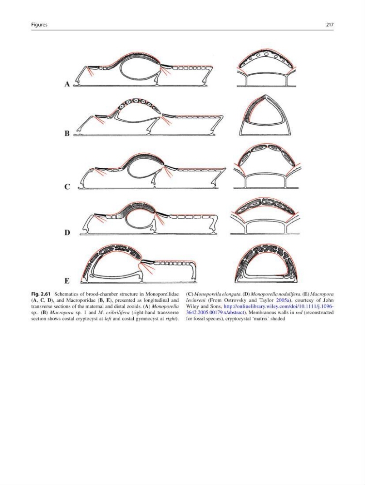
Fig. 2.61 Schematics of brood-chamber structure in Monoporellidae
( A , C , D ), and Macroporidae ( B , E ), presented as longitudinal and
transverse sections of the maternal and distal zooids. ( A ) Monoporella
sp.. ( B ) Macropora sp. 1 and M . cribrilifera (right-hand transverse
section shows costal cryptocyst at left and costal gymnocyst at right ).
( C ) Monoporella elongata . ( D ) Monoporella nodulifera . ( E ) Macropora
levinseni (From Ostrovsky and Taylor 2005a ), courtesy of John
Wiley and Sons, http://onlinelibrary.wiley.com/doi/10.1111/j.10963642.2005.00179.x/abstract ). Membranous walls in red (reconstructed
for fossil species), cryptocystal ‘matrix’ shaded
Figures
Fig. 2.61 Schematics of brood-chamber structure in Monoporellidae
( A , C , D ), and Macroporidae ( B , E ), presented as longitudinal and
transverse sections of the maternal and distal zooids. ( A ) Monoporella
sp.. ( B ) Macropora sp. 1 and M . cribrilifera (right-hand transverse
section shows costal cryptocyst at left and costal gymnocyst at right ).
( C ) Monoporella elongata . ( D ) Monoporella nodulifera . ( E ) Macropora
levinseni (From Ostrovsky and Taylor 2005a ), courtesy of John
Wiley and Sons, http://onlinelibrary.wiley.com/doi/10.1111/j.10963642.2005.00179.x/abstract ). Membranous walls in red (reconstructed
for fossil species), cryptocystal ‘matrix’ shaded
Figures