

213
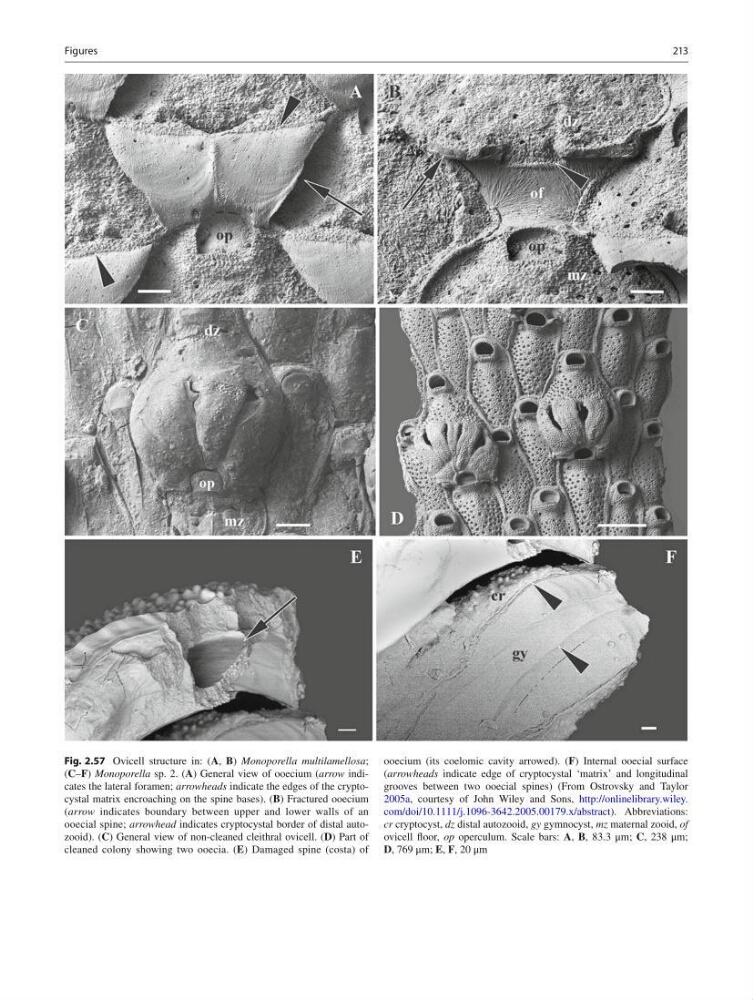
Fig. 2.57 Ovicell structure in: ( A , B ) Monoporella multilamellosa ;
( C – F ) Monoporella sp. 2. ( A ) General view of ooecium ( arrow indicates the lateral foramen; arrowheads indicate the edges of the cryptocystal matrix encroaching on the spine bases). ( B ) Fractured ooecium
( arrow indicates boundary between upper and lower walls of an
ooecial spine; arrowhead indicates cryptocystal border of distal autozooid). ( C ) General view of non-cleaned cleithral ovicell. ( D ) Part of
cleaned colony showing two ooecia. ( E ) Damaged spine (costa) of
ooecium (its coelomic cavity arrowed). ( F ) Internal ooecial surface
( arrowheads indicate edge of cryptocystal ‘matrix’ and longitudinal
grooves between two ooecial spines) (From Ostrovsky and Taylor
2005a , courtesy of John Wiley and Sons, http://onlinelibrary.wiley.
com/doi/10.1111/j.1096-3642.2005.00179.x/abstract ). Abbreviations:
cr cryptocyst, dz distal autozooid, gy gymnocyst, mz maternal zooid, of
ovicell fl oor, op operculum. Scale bars: A , B , 83.3 μm; C , 238 μm;
D , 769 μm; E , F , 20 μm
Figures
Fig. 2.57 Ovicell structure in: ( A , B ) Monoporella multilamellosa ;
( C – F ) Monoporella sp. 2. ( A ) General view of ooecium ( arrow indicates the lateral foramen; arrowheads indicate the edges of the cryptocystal matrix encroaching on the spine bases). ( B ) Fractured ooecium
( arrow indicates boundary between upper and lower walls of an
ooecial spine; arrowhead indicates cryptocystal border of distal autozooid). ( C ) General view of non-cleaned cleithral ovicell. ( D ) Part of
cleaned colony showing two ooecia. ( E ) Damaged spine (costa) of
ooecium (its coelomic cavity arrowed). ( F ) Internal ooecial surface
( arrowheads indicate edge of cryptocystal ‘matrix’ and longitudinal
grooves between two ooecial spines) (From Ostrovsky and Taylor
2005a , courtesy of John Wiley and Sons, http://onlinelibrary.wiley.
com/doi/10.1111/j.1096-3642.2005.00179.x/abstract ). Abbreviations:
cr cryptocyst, dz distal autozooid, gy gymnocyst, mz maternal zooid, of
ovicell fl oor, op operculum. Scale bars: A , B , 83.3 μm; C , 238 μm;
D , 769 μm; E , F , 20 μm
Figures