

191
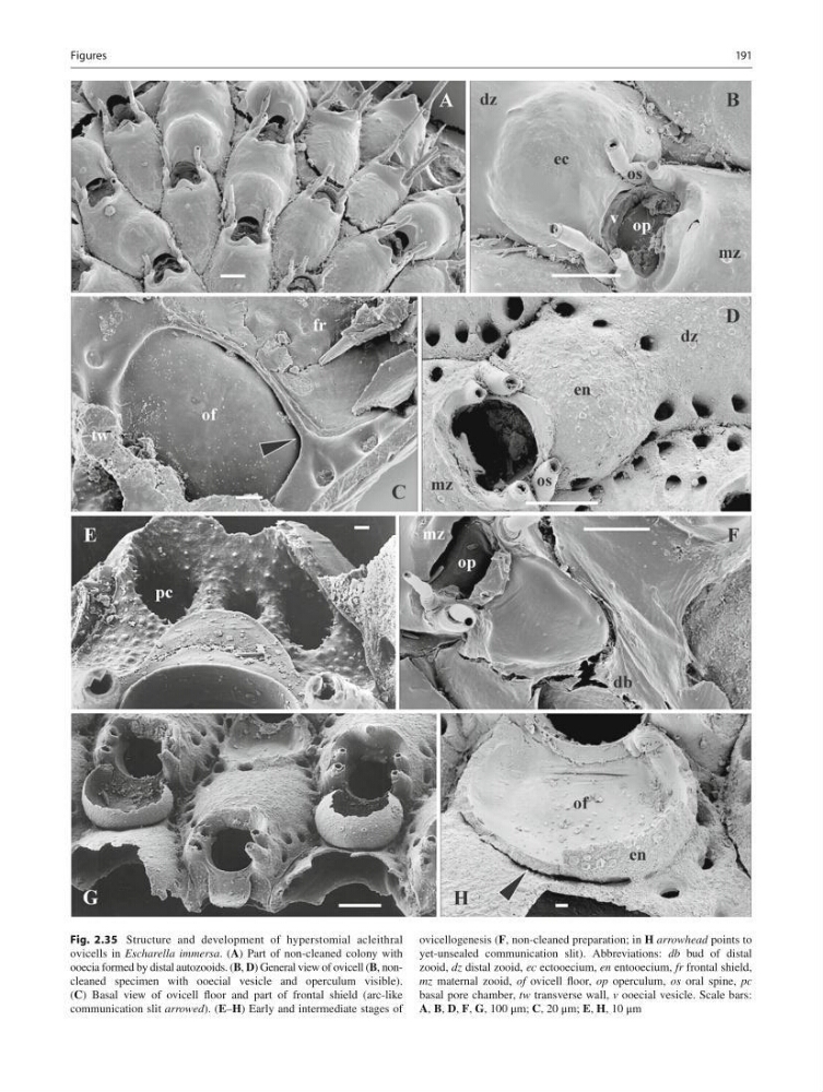
Fig. 2.35 Structure and development of hyperstomial acleithral
ovicells in Escharella immersa . ( A ) Part of non-cleaned colony with
ooecia formed by distal autozooids. ( B , D ) General view of ovicell ( B , noncleaned specimen with ooecial vesicle and operculum visible).
( C ) Basal view of ovicell fl oor and part of frontal shield (arc-like
communication slit arrowed ). ( E – H ) Early and intermediate stages of
ovicellogenesis ( F , non- cleaned preparation; in H arrowhead points to
yet-unsealed communication slit). Abbreviations: db bud of distal
zooid, dz distal zooid, ес ectooecium, en entooecium, fr frontal shield,
mz maternal zooid, of ovicell fl oor, op operculum, os oral spine, рс
basal pore chamber, tw transverse wall, v ooecial vesicle. Scale bars:
A , B , D , F , G , 100 μm; C , 20 μm; E , H , 10 μm
Figures
Fig. 2.35 Structure and development of hyperstomial acleithral
ovicells in Escharella immersa . ( A ) Part of non-cleaned colony with
ooecia formed by distal autozooids. ( B , D ) General view of ovicell ( B , noncleaned specimen with ooecial vesicle and operculum visible).
( C ) Basal view of ovicell fl oor and part of frontal shield (arc-like
communication slit arrowed ). ( E – H ) Early and intermediate stages of
ovicellogenesis ( F , non- cleaned preparation; in H arrowhead points to
yet-unsealed communication slit). Abbreviations: db bud of distal
zooid, dz distal zooid, ес ectooecium, en entooecium, fr frontal shield,
mz maternal zooid, of ovicell fl oor, op operculum, os oral spine, рс
basal pore chamber, tw transverse wall, v ooecial vesicle. Scale bars:
A , B , D , F , G , 100 μm; C , 20 μm; E , H , 10 μm
Figures