

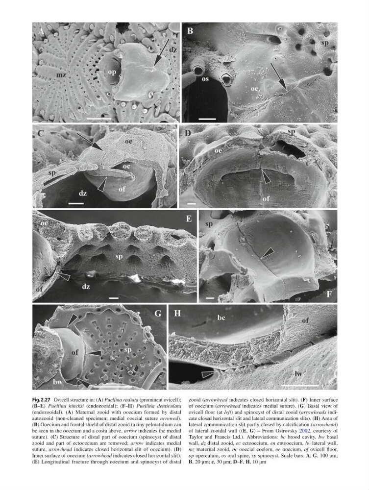
Fig. 2.27 Ovicell structure in: ( A ) Puellina radiata (prominent ovicell);
( B – E ) Puellina hincksi (endozooidal); ( F – H ) Puellina denticulata
(endozooidal). ( A ) Maternal zooid with ooecium formed by distal
autozooid (non-cleaned specimen; medial ooecial suture arrowed ).
( B ) Ooecium and frontal shield of distal zooid (a tiny pelmatidium can
be seen in the ooecium and a costa above, arrow indicates the medial
suture). ( C ) Structure of distal part of ooecium (spinocyst of distal
zooid and part of ectooecium are removed; arrow indicates medial
suture, arrowhead indicates closed horizontal slit of ooecium). ( D )
Inner surface of ooecium ( arrowhead indicates closed horizontal slit).
( E ) Longitudinal fracture through ooecium and spinocyst of distal
zooid ( arrowhead indicates closed horizontal slit). ( F ) Inner surface
of ooecium ( arrowhead indicates medial suture). ( G ) Basal view of
ovicell fl oor (at left ) and spinocyst of distal zooid ( arrowheads indicate closed horizontal slit and lateral communication slits). ( H ) Area of
lateral communication slit partly closed by calcifi cation ( arrowhead )
of lateral zooidal wall (( E , G ) – From Ostrovsky 2002 , courtesy of
Taylor and Francis Ltd.). Abbreviations: bc brood cavity, bw basal
wall, dz distal zooid, ес ectooecium, en entooecium, lw lateral wall,
mz maternal zooid, oc ooecial coelom, oe ooecium, of ovicell fl oor,
op operculum, os oral spine, sp spinocyst. Scale bars: A , G , 100 μm;
B , 20 μm; c , 30 μm; D – F , H , 10 μm
( B – E ) Puellina hincksi (endozooidal); ( F – H ) Puellina denticulata
(endozooidal). ( A ) Maternal zooid with ooecium formed by distal
autozooid (non-cleaned specimen; medial ooecial suture arrowed ).
( B ) Ooecium and frontal shield of distal zooid (a tiny pelmatidium can
be seen in the ooecium and a costa above, arrow indicates the medial
suture). ( C ) Structure of distal part of ooecium (spinocyst of distal
zooid and part of ectooecium are removed; arrow indicates medial
suture, arrowhead indicates closed horizontal slit of ooecium). ( D )
Inner surface of ooecium ( arrowhead indicates closed horizontal slit).
( E ) Longitudinal fracture through ooecium and spinocyst of distal
zooid ( arrowhead indicates closed horizontal slit). ( F ) Inner surface
of ooecium ( arrowhead indicates medial suture). ( G ) Basal view of
ovicell fl oor (at left ) and spinocyst of distal zooid ( arrowheads indicate closed horizontal slit and lateral communication slits). ( H ) Area of
lateral communication slit partly closed by calcifi cation ( arrowhead )
of lateral zooidal wall (( E , G ) – From Ostrovsky 2002 , courtesy of
Taylor and Francis Ltd.). Abbreviations: bc brood cavity, bw basal
wall, dz distal zooid, ес ectooecium, en entooecium, lw lateral wall,
mz maternal zooid, oc ooecial coelom, oe ooecium, of ovicell fl oor,
op operculum, os oral spine, sp spinocyst. Scale bars: A , G , 100 μm;
B , 20 μm; c , 30 μm; D – F , H , 10 μm