

173
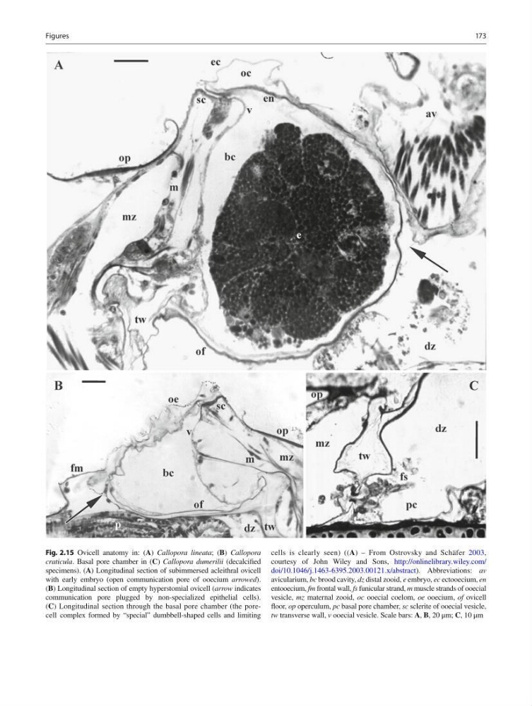
Fig. 2.15 Ovicell anatomy in: ( A ) Callopora lineata ; ( B ) Callopora
craticula . Basal pore chamber in ( C ) Callopora dumerilii (decalcifi ed
specimens). ( A ) Longitudinal section of subimmersed acleithral ovicell
with early embryo (open communication pore of ooecium arrowed ).
( B ) Longitudinal section of empty hyperstomial ovicell ( arrow indicates
communication pore plugged by non-specialized epithelial cells).
( C ) Longitudinal section through the basal pore chamber (the porecell complex formed by “special” dumbbell-shaped cells and limiting
cells is clearly seen) (( A ) – From Ostrovsky and Schäfer 2003 ,
courtesy of John Wiley and Sons, http://onlinelibrary.wiley.com/
doi/10.1046/j.1463-6395.2003.00121.x/abstract ). Abbreviations: av
avicularium, bc brood cavity, dz distal zooid, е embryo, ес ectooecium, en
entooecium, fm frontal wall, fs funicular strand, m muscle strands of ooecial
vesicle, mz maternal zooid, oc ooecial coelom, ое ooecium, of ovicell
fl oor, op operculum, pc basal pore chamber, sc sclerite of ooecial vesicle,
tw transverse wall, v ooecial vesicle. Scale bars: A , B , 20 μm; C , 10 μm
Figures
Fig. 2.15 Ovicell anatomy in: ( A ) Callopora lineata ; ( B ) Callopora
craticula . Basal pore chamber in ( C ) Callopora dumerilii (decalcifi ed
specimens). ( A ) Longitudinal section of subimmersed acleithral ovicell
with early embryo (open communication pore of ooecium arrowed ).
( B ) Longitudinal section of empty hyperstomial ovicell ( arrow indicates
communication pore plugged by non-specialized epithelial cells).
( C ) Longitudinal section through the basal pore chamber (the porecell complex formed by “special” dumbbell-shaped cells and limiting
cells is clearly seen) (( A ) – From Ostrovsky and Schäfer 2003 ,
courtesy of John Wiley and Sons, http://onlinelibrary.wiley.com/
doi/10.1046/j.1463-6395.2003.00121.x/abstract ). Abbreviations: av
avicularium, bc brood cavity, dz distal zooid, е embryo, ес ectooecium, en
entooecium, fm frontal wall, fs funicular strand, m muscle strands of ooecial
vesicle, mz maternal zooid, oc ooecial coelom, ое ooecium, of ovicell
fl oor, op operculum, pc basal pore chamber, sc sclerite of ooecial vesicle,
tw transverse wall, v ooecial vesicle. Scale bars: A , B , 20 μm; C , 10 μm
Figures