

131
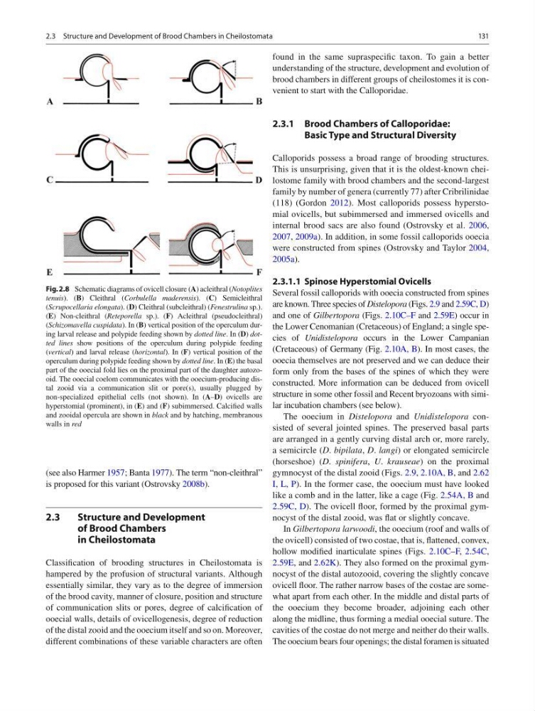
(see also Harmer 1957 ; Banta 1977 ). The term “non- cleithral”
is proposed for this variant (Ostrovsky 2008b ).
2.3
Structure and Development
of Brood Chambers
in Cheilostomata
Classifi cation of brooding structures in Cheilostomata is
hampered by the profusion of structural variants. Although
essentially similar, they vary as to the degree of immersion
of the brood cavity, manner of closure, position and structure
of communication slits or pores, degree of calcifi cation of
ooecial walls, details of ovicellogenesis, degree of reduction
of the distal zooid and the ooecium itself and so on. Moreover,
different combinations of these variable characters are often
found in the same supraspecifi c taxon. To gain a better
understanding of the structure, development and evolution of
brood chambers in different groups of cheilostomes it is convenient to start with the Calloporidae.
2.3.1 Brood Chambers of Calloporidae:
Basic Type and Structural Diversity
Calloporids possess a broad range of brooding structures.
This is unsurprising, given that it is the oldest-known cheilostome family with brood chambers and the second-largest
family by number of genera (currently 77) after Cribrilinidae
(118) (Gordon 2012 ). Most calloporids possess hyperstomial ovicells, but subimmersed and immersed ovicells and
internal brood sacs are also found (Ostrovsky et al. 2006 ,
2007 , 2009a ). In addition, in some fossil calloporids ooecia
were constructed from spines (Ostrovsky and Taylor 2004 ,
2005a ).
2.3.1.1 Spinose Hyperstomial Ovicells
Several fossil calloporids with ooecia constructed from spines
are known. Three species of Distelopora (Figs. 2.9 and 2.59C, D )
and one of Gilbertopora (Figs. 2.10C–F and 2.59E ) occur in
the Lower Cenomanian (Cretaceous) of England; a single species of Unidistelopora occurs in the Lower Campanian
(Cretaceous) of Germany (Fig. 2.10A, B ). In most cases, the
ooecia themselves are not preserved and we can deduce their
form only from the bases of the spines of which they were
constructed. More information can be deduced from ovicell
structure in some other fossil and Recent bryozoans with similar incubation chambers (see below).
The ooecium in Distelopora and Unidistelopora consisted of several jointed spines. The preserved basal parts
are arranged in a gently curving distal arch or, more rarely,
a semicircle ( D . bipilata , D . langi ) or elongated semicircle
(horseshoe) ( D . spinifera , U . krauseae ) on the proximal
gymnocyst of the distal zooid (Figs. 2.9 , 2.10A, B , and 2.62
I, L, P ). In the former case, the ooecium must have looked
like a comb and in the latter, like a cage (Fig. 2.54A, B and
2.59C, D ). The ovicell fl oor, formed by the proximal gymnocyst of the distal zooid, was fl at or slightly concave.
In Gilbertopora larwoodi , the ooecium (roof and walls of
the ovicell) consisted of two costae, that is, fl attened, convex,
hollow modifi ed inarticulate spines (Figs. 2.10C–F , 2.54C ,
2.59E , and 2.62K ). They also formed on the proximal gymnocyst of the distal autozooid, covering the slightly concave
ovicell fl oor. The rather narrow bases of the costae are somewhat apart from each other. In the middle and distal parts of
the ooecium they become broader, adjoining each other
along the midline, thus forming a medial ooecial suture. The
cavities of the costae do not merge and neither do their walls.
The ooecium bears four openings; the distal foramen is situated
Fig. 2.8 Schematic diagrams of ovicell closure ( A ) acleithral ( Notoplites
tenuis ). ( B ) Cleithral ( Corbulella maderensis ). ( C ) Semicleithral
( Scrupocellaria elongata ). ( D ) Cleithral (subcleithral) ( Fenestrulina sp.).
( E ) Non-cleithral ( Reteporella sp.). ( F ) Acleithral (pseudocleithral)
( Schizomavella cuspidata ). In ( B ) vertical position of the operculum during larval release and polypide feeding shown by dotted line . In ( D ) dotted lines show positions of the operculum during polypide feeding
( vertical ) and larval release ( horizontal ). In ( F ) vertical position of the
operculum during polypide feeding shown by dotted line . In ( E ) the basal
part of the ooecial fold lies on the proximal part of the daughter autozooid. The ooecial coelom communicates with the ooecium-producing distal zooid via a communication slit or pore(s), usually plugged by
non-specialized epithelial cells (not shown). In ( A – D ) ovicells are
hyperstomial (prominent), in ( E ) and ( F ) subimmersed. Calcifi ed walls
and zooidal opercula are shown in black and by hatching, membranous
walls in red
2.3 Structure and Development of Brood Chambers in Cheilostomata
(see also Harmer 1957 ; Banta 1977 ). The term “non- cleithral”
is proposed for this variant (Ostrovsky 2008b ).
2.3
Structure and Development
of Brood Chambers
in Cheilostomata
Classifi cation of brooding structures in Cheilostomata is
hampered by the profusion of structural variants. Although
essentially similar, they vary as to the degree of immersion
of the brood cavity, manner of closure, position and structure
of communication slits or pores, degree of calcifi cation of
ooecial walls, details of ovicellogenesis, degree of reduction
of the distal zooid and the ooecium itself and so on. Moreover,
different combinations of these variable characters are often
found in the same supraspecifi c taxon. To gain a better
understanding of the structure, development and evolution of
brood chambers in different groups of cheilostomes it is convenient to start with the Calloporidae.
2.3.1 Brood Chambers of Calloporidae:
Basic Type and Structural Diversity
Calloporids possess a broad range of brooding structures.
This is unsurprising, given that it is the oldest-known cheilostome family with brood chambers and the second-largest
family by number of genera (currently 77) after Cribrilinidae
(118) (Gordon 2012 ). Most calloporids possess hyperstomial ovicells, but subimmersed and immersed ovicells and
internal brood sacs are also found (Ostrovsky et al. 2006 ,
2007 , 2009a ). In addition, in some fossil calloporids ooecia
were constructed from spines (Ostrovsky and Taylor 2004 ,
2005a ).
2.3.1.1 Spinose Hyperstomial Ovicells
Several fossil calloporids with ooecia constructed from spines
are known. Three species of Distelopora (Figs. 2.9 and 2.59C, D )
and one of Gilbertopora (Figs. 2.10C–F and 2.59E ) occur in
the Lower Cenomanian (Cretaceous) of England; a single species of Unidistelopora occurs in the Lower Campanian
(Cretaceous) of Germany (Fig. 2.10A, B ). In most cases, the
ooecia themselves are not preserved and we can deduce their
form only from the bases of the spines of which they were
constructed. More information can be deduced from ovicell
structure in some other fossil and Recent bryozoans with similar incubation chambers (see below).
The ooecium in Distelopora and Unidistelopora consisted of several jointed spines. The preserved basal parts
are arranged in a gently curving distal arch or, more rarely,
a semicircle ( D . bipilata , D . langi ) or elongated semicircle
(horseshoe) ( D . spinifera , U . krauseae ) on the proximal
gymnocyst of the distal zooid (Figs. 2.9 , 2.10A, B , and 2.62
I, L, P ). In the former case, the ooecium must have looked
like a comb and in the latter, like a cage (Fig. 2.54A, B and
2.59C, D ). The ovicell fl oor, formed by the proximal gymnocyst of the distal zooid, was fl at or slightly concave.
In Gilbertopora larwoodi , the ooecium (roof and walls of
the ovicell) consisted of two costae, that is, fl attened, convex,
hollow modifi ed inarticulate spines (Figs. 2.10C–F , 2.54C ,
2.59E , and 2.62K ). They also formed on the proximal gymnocyst of the distal autozooid, covering the slightly concave
ovicell fl oor. The rather narrow bases of the costae are somewhat apart from each other. In the middle and distal parts of
the ooecium they become broader, adjoining each other
along the midline, thus forming a medial ooecial suture. The
cavities of the costae do not merge and neither do their walls.
The ooecium bears four openings; the distal foramen is situated
Fig. 2.8 Schematic diagrams of ovicell closure ( A ) acleithral ( Notoplites
tenuis ). ( B ) Cleithral ( Corbulella maderensis ). ( C ) Semicleithral
( Scrupocellaria elongata ). ( D ) Cleithral (subcleithral) ( Fenestrulina sp.).
( E ) Non-cleithral ( Reteporella sp.). ( F ) Acleithral (pseudocleithral)
( Schizomavella cuspidata ). In ( B ) vertical position of the operculum during larval release and polypide feeding shown by dotted line . In ( D ) dotted lines show positions of the operculum during polypide feeding
( vertical ) and larval release ( horizontal ). In ( F ) vertical position of the
operculum during polypide feeding shown by dotted line . In ( E ) the basal
part of the ooecial fold lies on the proximal part of the daughter autozooid. The ooecial coelom communicates with the ooecium-producing distal zooid via a communication slit or pore(s), usually plugged by
non-specialized epithelial cells (not shown). In ( A – D ) ovicells are
hyperstomial (prominent), in ( E ) and ( F ) subimmersed. Calcifi ed walls
and zooidal opercula are shown in black and by hatching, membranous
walls in red
2.3 Structure and Development of Brood Chambers in Cheilostomata