

126
distinct frontal part (see also Ristedt 1985 ; Harmelin and
Arístegui 1988 ). The ooecium itself is the frontal or distal
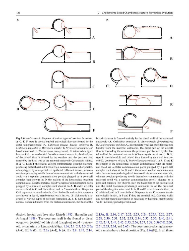
outgrowth (outfold) of this distal (daughter) zooid – autozooid, avicularium or kenozooid (Figs. 1.36 , 2.1 , 2.3 , 2.5 , 2.6a
(A–C, E), b (D, F), 2.7a (A–I), b (A, B), 2.8 , 2.13 , 2.14 ,
2.15A, B , 2.16 , 2.17 , 2.22 , 2.23 , 2.24 , 2.25A , 2.26 , 2.27 ,
2.28 , 2.30 , 2.31 , 2.32 , 2.33 , 2.34 , 2.35 , 2.36 , 2.40 , 2.41 ,
2.42 , 2.43 , 2.44 , 2.45 , 2.55 , 2.56 , 2.57 , 2.58 , 2.59C-E , 2.60 ,
2.61 , 2.63 , 2.64 , and 2.65 ). The ooecium-producing kenozooid can also have a basal position (Fig. 2.6a (F)). In all these
Fig. 2.6 ( a ) Schematic diagrams of various types of ooecium formation.
A – C , E , F , type 1: ooecial outfold and ovicell fl oor are formed by the
distal (auto/keno)zooid ( A , Callopora lineata , Tegella armifera ; B ,
Callopora dumerilii ; C , Micropora notialis ; E , Bryocalyx cinnameus ), or
basal kenozooid ( F , Cornucopina pectogemma ). D , intermediate type:
kenozooidal ooecium budded from the maternal autozooid, the distal part
of the ovicell fl oor is formed by the ooecium and the proximal part
formed by the distal wall of the maternal autozooid ( Costaticella solida ).
In A – C , E and F the ooecial coelom communicates with the ooeciumproducing distal (basal in F ) zooid via a communication slit or pore(s),
often plugged by non-specialized epithelial cells (not shown), whereas
ooecium-producing zooids themselves communicate with the maternal
zooid via a septular communication pore(s) plugged by a pore-cell
complex (not shown). In D , the coelom of the kenozooidal ooecium
communicates with the maternal zooid via septular communication pores
plugged by a pore-cell complex (not shown). In A , B and E ovicells
are acleithral, in C and D cleithral, and in F semicleithral. Diagrams
C – F represent terminal ovicells. Calcifi ed walls and zooidal opercula
are shown in black , membranous walls in red . ( b ) Schematic diagrams of various types of ooecium formation. A , B , E , type 2: kenozooidal ooecium budded from the maternal autozooid, the fl oor of the
brood chamber is formed entirely by the distal wall of the maternal
autozooid ( A , Cribrilina annulata ; B , Eurystomella foraminigera ;
E , Cauloramphus spinifer ). C , intermediate type: kenozooidal ooecium
budded from the maternal autozooid, the distal part of the ovicell
fl oor is formed by the ooecium, the proximal part formed by the distal wall of the maternal autozooid ( Chaperiopsis cervicornis ). D , F ,
type 1: ooecial outfold and ovicell fl oor formed by the distal kenozooid ( D , Omanipora pilleri ; F , Turbicellepora crenulata ). In A – C and E
the coelom of the kenozooidal ooecium communicates with the maternal zooid via septular communication pores plugged by a pore-cell
complex (not shown). In D and F the ooecial coelom communicates
with the ooecium-producing distal kenozooid via a communication slit,
whereas ooecium- producing zooids themselves communicate with the
maternal zooid via a septular communication pore(s) plugged by a
pore-cell complex (not shown). In F the basal part of the ooecial fold
and the distal (ooecium-producing) kenozooid lie on the proximal
part of the daughter autozooid. In A , B and D ovicells are cleithral, in
C acleithral, and in F non-cleithral. Diagrams A and C represent terminal ovicells (in fact, in B and F they are terminal too). Calcifi ed walls
and zooidal opercula are shown in black and by hatching, membranous
walls (including pseudopores) in red
2 Cheilostome Brood Chambers: Structure, Formation, Evolution
distinct frontal part (see also Ristedt 1985 ; Harmelin and
Arístegui 1988 ). The ooecium itself is the frontal or distal
outgrowth (outfold) of this distal (daughter) zooid – autozooid, avicularium or kenozooid (Figs. 1.36 , 2.1 , 2.3 , 2.5 , 2.6a
(A–C, E), b (D, F), 2.7a (A–I), b (A, B), 2.8 , 2.13 , 2.14 ,
2.15A, B , 2.16 , 2.17 , 2.22 , 2.23 , 2.24 , 2.25A , 2.26 , 2.27 ,
2.28 , 2.30 , 2.31 , 2.32 , 2.33 , 2.34 , 2.35 , 2.36 , 2.40 , 2.41 ,
2.42 , 2.43 , 2.44 , 2.45 , 2.55 , 2.56 , 2.57 , 2.58 , 2.59C-E , 2.60 ,
2.61 , 2.63 , 2.64 , and 2.65 ). The ooecium-producing kenozooid can also have a basal position (Fig. 2.6a (F)). In all these
Fig. 2.6 ( a ) Schematic diagrams of various types of ooecium formation.
A – C , E , F , type 1: ooecial outfold and ovicell fl oor are formed by the
distal (auto/keno)zooid ( A , Callopora lineata , Tegella armifera ; B ,
Callopora dumerilii ; C , Micropora notialis ; E , Bryocalyx cinnameus ), or
basal kenozooid ( F , Cornucopina pectogemma ). D , intermediate type:
kenozooidal ooecium budded from the maternal autozooid, the distal part
of the ovicell fl oor is formed by the ooecium and the proximal part
formed by the distal wall of the maternal autozooid ( Costaticella solida ).
In A – C , E and F the ooecial coelom communicates with the ooeciumproducing distal (basal in F ) zooid via a communication slit or pore(s),
often plugged by non-specialized epithelial cells (not shown), whereas
ooecium-producing zooids themselves communicate with the maternal
zooid via a septular communication pore(s) plugged by a pore-cell
complex (not shown). In D , the coelom of the kenozooidal ooecium
communicates with the maternal zooid via septular communication pores
plugged by a pore-cell complex (not shown). In A , B and E ovicells
are acleithral, in C and D cleithral, and in F semicleithral. Diagrams
C – F represent terminal ovicells. Calcifi ed walls and zooidal opercula
are shown in black , membranous walls in red . ( b ) Schematic diagrams of various types of ooecium formation. A , B , E , type 2: kenozooidal ooecium budded from the maternal autozooid, the fl oor of the
brood chamber is formed entirely by the distal wall of the maternal
autozooid ( A , Cribrilina annulata ; B , Eurystomella foraminigera ;
E , Cauloramphus spinifer ). C , intermediate type: kenozooidal ooecium
budded from the maternal autozooid, the distal part of the ovicell
fl oor is formed by the ooecium, the proximal part formed by the distal wall of the maternal autozooid ( Chaperiopsis cervicornis ). D , F ,
type 1: ooecial outfold and ovicell fl oor formed by the distal kenozooid ( D , Omanipora pilleri ; F , Turbicellepora crenulata ). In A – C and E
the coelom of the kenozooidal ooecium communicates with the maternal zooid via septular communication pores plugged by a pore-cell
complex (not shown). In D and F the ooecial coelom communicates
with the ooecium-producing distal kenozooid via a communication slit,
whereas ooecium- producing zooids themselves communicate with the
maternal zooid via a septular communication pore(s) plugged by a
pore-cell complex (not shown). In F the basal part of the ooecial fold
and the distal (ooecium-producing) kenozooid lie on the proximal
part of the daughter autozooid. In A , B and D ovicells are cleithral, in
C acleithral, and in F non-cleithral. Diagrams A and C represent terminal ovicells (in fact, in B and F they are terminal too). Calcifi ed walls
and zooidal opercula are shown in black and by hatching, membranous
walls (including pseudopores) in red
2 Cheilostome Brood Chambers: Structure, Formation, Evolution