

122
( 1900 ) has often been omitted without comment (Hyman
1959 ; Brien 1960 ; Prenant and Bobin 1966 ). This may have
happened because Calvet stressed that he did not fi nd this
pore in any of the species with hyperstomial ovicells other
than Bugula simplex , even though the connection between
ooecial and visceral coeloms was described and depicted
by him in the endozooidal ovicell of Securifl ustra securifrons (see Calvet 1900 , fi g. 44).
Other than Hass ( 1948 , fi g. 32), who correctly depicted
the lumen of the ooecial fold connected with the visceral
coelom of the distal zooid via “Oözialporus” in a phidoloporid
cheilostome (as Sertella ), no-one challenged Silén’s generally accepted opinion during the next three decades. Ryland
( 1962 , 1965 , 1968 ) and Moyano ( 1968 ) depicted ooecia
either resting on the frontal wall of the distal zooid or
immersed in it, but gave no details of their communication
with the visceral coelom. In the latter work, Ryland ( 1968 )
discussed terminological problems subsequent to the works
of Jullien ( 1888 ), Levinsen ( 1902 , 1909 ), Canu and Bassler
( 1920 ), and Silén ( 1944 , 1945 ) and selected the most appropriate terms that are currently in use (see also Ryland 1976 ,
1982 ; reviewed in Ostrovsky 2008a ).
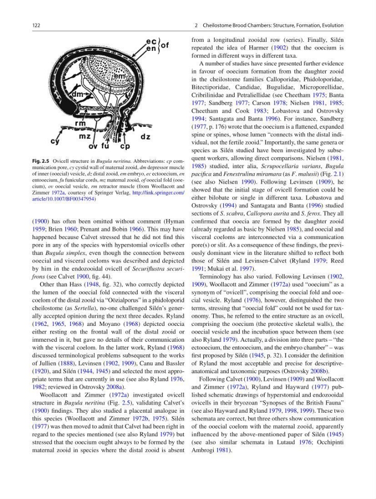
Woollacott and Zimmer ( 1972a ) investigated ovicell
structure in Bugula neritina (Fig. 2.5 ), validating Calvet’s
( 1900 ) fi ndings. They also studied a placental analogue in
this species (Woollacott and Zimmer 1972b , 1975 ). Silén
( 1977 ) was then moved to admit that Calvet had been right in
regard to the species mentioned (see also Ryland 1979 ) but
stressed that the ooecium ought always to be formed by the
maternal zooid in species where the distal zooid is absent
from a longitudinal zooidal row (series). Finally, Silén
repeated the idea of Harmer ( 1902 ) that the ooecium is
formed in different ways in different taxa.
A number of studies have since presented further evidence
in favour of ooecium formation from the daughter zooid
in the cheilostome families Calloporidae, Phidoloporidae,
Bitectiporidae, Candidae, Bugulidae, Microporellidae,
Cribrilinidae and Petraliellidae (see Cheetham 1975 ; Banta
1977 ; Sandberg 1977 ; Carson 1978 ; Nielsen 1981 , 1985 ;
Cheetham and Cook 1983 ; Lobastova and Ostrovsky
1994 ; Santagata and Banta 1996 ). For instance, Sandberg
( 1977 , p. 176) wrote that the ooecium is a fl attened, expanded
spine or spines, whose lumen “connects with the distal individual, not the fertile zooid.” Importantly, the same genera or
species as Silén studied have been investigated by subsequent workers, allowing direct comparisons. Nielsen ( 1981 ,
1985 ) studied, inter alia, Scrupocellaria varians , Bugula
pacifi ca and Fenestrulina miramara (as F . malusii ) (Fig. 2.1 )
(see also Nielsen 1990 ). Following Levinsen ( 1909 ), he
showed that the initial stage of ovicell formation could be
either bilobate or single in different taxa. Lobastova and
Ostrovsky ( 1994 ) and Santagata and Banta ( 1996 ) studied
sections of S . scabra , Callopora aurita and S . ferox . They all
confi rmed that ooecia are formed by the daughter zooid
(already regarded as basic by Nielsen 1985 ), and ooecial and
visceral coeloms are interconnected via a communication
pore(s) or slit. As a consequence of these fi ndings, the previously dominant view in the literature shifted to refl ect both
those of Silén and Levinsen-Calvet (Ryland 1979 ; Reed
1991 ; Mukai et al. 1997 ).
Terminology has also varied. Following Levinsen ( 1902 ,
1909 ), Woollacott and Zimmer ( 1972a ) used “ooecium” as a
synonym of “ovicell”, comprising the ooecial fold and ooecial vesicle. Ryland ( 1976 ), however, distinguished the two
terms, stressing that “ooecial fold” could not be used for taxonomy. Thus, he referred to the entire structure as an ovicell,
comprising the ooecium (the protective skeletal walls), the
ooecial vesicle and the incubation space between them (see
also Ryland 1979 ). Actually, a division into three parts – “the
ectooecium, the entooecium, and the embryo chamber” – was
fi rst proposed by Silén ( 1945 , p. 32). I consider the defi nition
of Ryland the most acceptable and precise for descriptiveanatomical and taxonomic purposes (Ostrovsky 2008b ).
Following Calvet ( 1900 ), Levinsen ( 1909 ) and Woollacott
and Zimmer ( 1972a ), Ryland and Hayward ( 1977 ) published schematic drawings of hyperstomial and endozooidal
ovicells in their bryozoan “Synopses of the British Fauna”
(see also Hayward and Ryland 1979 , 1998 , 1999 ). These two
schemata are correct, but three others show communication
of the ooecial coelom with the maternal zooid, apparently
infl uenced by the above-mentioned paper of Silén ( 1945 )
(see also similar schemata in Lutaud 1976 ; Occhipinti
Ambrogi 1981 ).
Fig. 2.5 Ovicell structure in Bugula neritina. Abbreviations: cp communication pore, cy cystid wall of maternal zooid, dm depressor muscle
of inner (ooecial) vesicle, dz distal zooid, em embryo, ec ectooecium, en
entooecium, fu funicular cords, mz maternal zooid, of ooecial fold (ooecium), ov ooecial vesicle, rm retractor muscle (from Woollacott and
Zimmer 1972a , courtesy of Springer Verlag, http://link.springer.com/
article/10.1007/BF00347954 )
2 Cheilostome Brood Chambers: Structure, Formation, Evolution
( 1900 ) has often been omitted without comment (Hyman
1959 ; Brien 1960 ; Prenant and Bobin 1966 ). This may have
happened because Calvet stressed that he did not fi nd this
pore in any of the species with hyperstomial ovicells other
than Bugula simplex , even though the connection between
ooecial and visceral coeloms was described and depicted
by him in the endozooidal ovicell of Securifl ustra securifrons (see Calvet 1900 , fi g. 44).
Other than Hass ( 1948 , fi g. 32), who correctly depicted
the lumen of the ooecial fold connected with the visceral
coelom of the distal zooid via “Oözialporus” in a phidoloporid
cheilostome (as Sertella ), no-one challenged Silén’s generally accepted opinion during the next three decades. Ryland
( 1962 , 1965 , 1968 ) and Moyano ( 1968 ) depicted ooecia
either resting on the frontal wall of the distal zooid or
immersed in it, but gave no details of their communication
with the visceral coelom. In the latter work, Ryland ( 1968 )
discussed terminological problems subsequent to the works
of Jullien ( 1888 ), Levinsen ( 1902 , 1909 ), Canu and Bassler
( 1920 ), and Silén ( 1944 , 1945 ) and selected the most appropriate terms that are currently in use (see also Ryland 1976 ,
1982 ; reviewed in Ostrovsky 2008a ).
Woollacott and Zimmer ( 1972a ) investigated ovicell
structure in Bugula neritina (Fig. 2.5 ), validating Calvet’s
( 1900 ) fi ndings. They also studied a placental analogue in
this species (Woollacott and Zimmer 1972b , 1975 ). Silén
( 1977 ) was then moved to admit that Calvet had been right in
regard to the species mentioned (see also Ryland 1979 ) but
stressed that the ooecium ought always to be formed by the
maternal zooid in species where the distal zooid is absent
from a longitudinal zooidal row (series). Finally, Silén
repeated the idea of Harmer ( 1902 ) that the ooecium is
formed in different ways in different taxa.
A number of studies have since presented further evidence
in favour of ooecium formation from the daughter zooid
in the cheilostome families Calloporidae, Phidoloporidae,
Bitectiporidae, Candidae, Bugulidae, Microporellidae,
Cribrilinidae and Petraliellidae (see Cheetham 1975 ; Banta
1977 ; Sandberg 1977 ; Carson 1978 ; Nielsen 1981 , 1985 ;
Cheetham and Cook 1983 ; Lobastova and Ostrovsky
1994 ; Santagata and Banta 1996 ). For instance, Sandberg
( 1977 , p. 176) wrote that the ooecium is a fl attened, expanded
spine or spines, whose lumen “connects with the distal individual, not the fertile zooid.” Importantly, the same genera or
species as Silén studied have been investigated by subsequent workers, allowing direct comparisons. Nielsen ( 1981 ,
1985 ) studied, inter alia, Scrupocellaria varians , Bugula
pacifi ca and Fenestrulina miramara (as F . malusii ) (Fig. 2.1 )
(see also Nielsen 1990 ). Following Levinsen ( 1909 ), he
showed that the initial stage of ovicell formation could be
either bilobate or single in different taxa. Lobastova and
Ostrovsky ( 1994 ) and Santagata and Banta ( 1996 ) studied
sections of S . scabra , Callopora aurita and S . ferox . They all
confi rmed that ooecia are formed by the daughter zooid
(already regarded as basic by Nielsen 1985 ), and ooecial and
visceral coeloms are interconnected via a communication
pore(s) or slit. As a consequence of these fi ndings, the previously dominant view in the literature shifted to refl ect both
those of Silén and Levinsen-Calvet (Ryland 1979 ; Reed
1991 ; Mukai et al. 1997 ).
Terminology has also varied. Following Levinsen ( 1902 ,
1909 ), Woollacott and Zimmer ( 1972a ) used “ooecium” as a
synonym of “ovicell”, comprising the ooecial fold and ooecial vesicle. Ryland ( 1976 ), however, distinguished the two
terms, stressing that “ooecial fold” could not be used for taxonomy. Thus, he referred to the entire structure as an ovicell,
comprising the ooecium (the protective skeletal walls), the
ooecial vesicle and the incubation space between them (see
also Ryland 1979 ). Actually, a division into three parts – “the
ectooecium, the entooecium, and the embryo chamber” – was
fi rst proposed by Silén ( 1945 , p. 32). I consider the defi nition
of Ryland the most acceptable and precise for descriptiveanatomical and taxonomic purposes (Ostrovsky 2008b ).
Following Calvet ( 1900 ), Levinsen ( 1909 ) and Woollacott
and Zimmer ( 1972a ), Ryland and Hayward ( 1977 ) published schematic drawings of hyperstomial and endozooidal
ovicells in their bryozoan “Synopses of the British Fauna”
(see also Hayward and Ryland 1979 , 1998 , 1999 ). These two
schemata are correct, but three others show communication
of the ooecial coelom with the maternal zooid, apparently
infl uenced by the above-mentioned paper of Silén ( 1945 )
(see also similar schemata in Lutaud 1976 ; Occhipinti
Ambrogi 1981 ).
Fig. 2.5 Ovicell structure in Bugula neritina. Abbreviations: cp communication pore, cy cystid wall of maternal zooid, dm depressor muscle
of inner (ooecial) vesicle, dz distal zooid, em embryo, ec ectooecium, en
entooecium, fu funicular cords, mz maternal zooid, of ooecial fold (ooecium), ov ooecial vesicle, rm retractor muscle (from Woollacott and
Zimmer 1972a , courtesy of Springer Verlag, http://link.springer.com/
article/10.1007/BF00347954 )
2 Cheilostome Brood Chambers: Structure, Formation, Evolution