

103
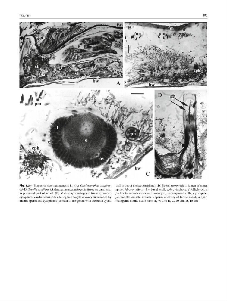
Fig. 1.34 Stages of spermatogenesis in: ( A ) Cauloramphus spinifer ;
( B – D ) Tegella armifera . ( A ) Immature spermatogenic tissue on basal wall
in proximal part of zooid. ( B ) Mature spermatogenic tissue (rounded
cytophores can be seen). ( C ) Vitellogenic oocyte in ovary surrounded by
mature sperm and cytophores (contact of the gonad with the basal cystid
wall is out of the section plane). ( D ) Sperm ( arrowed ) in lumen of mural
spine. Abbreviations: bw basal wall, cph cytophore, f follicle cells,
fm frontal membranous wall, o oocyte, ov ovary-wall cells, p polypide,
рm parietal muscle strands, s sperm in cavity of fertile zooid, st spermatogenic tissue. Scale bars: A , 40 μm; B , C , 20 μm; D , 10 μm
Figures
Fig. 1.34 Stages of spermatogenesis in: ( A ) Cauloramphus spinifer ;
( B – D ) Tegella armifera . ( A ) Immature spermatogenic tissue on basal wall
in proximal part of zooid. ( B ) Mature spermatogenic tissue (rounded
cytophores can be seen). ( C ) Vitellogenic oocyte in ovary surrounded by
mature sperm and cytophores (contact of the gonad with the basal cystid
wall is out of the section plane). ( D ) Sperm ( arrowed ) in lumen of mural
spine. Abbreviations: bw basal wall, cph cytophore, f follicle cells,
fm frontal membranous wall, o oocyte, ov ovary-wall cells, p polypide,
рm parietal muscle strands, s sperm in cavity of fertile zooid, st spermatogenic tissue. Scale bars: A , 40 μm; B , C , 20 μm; D , 10 μm
Figures