

96
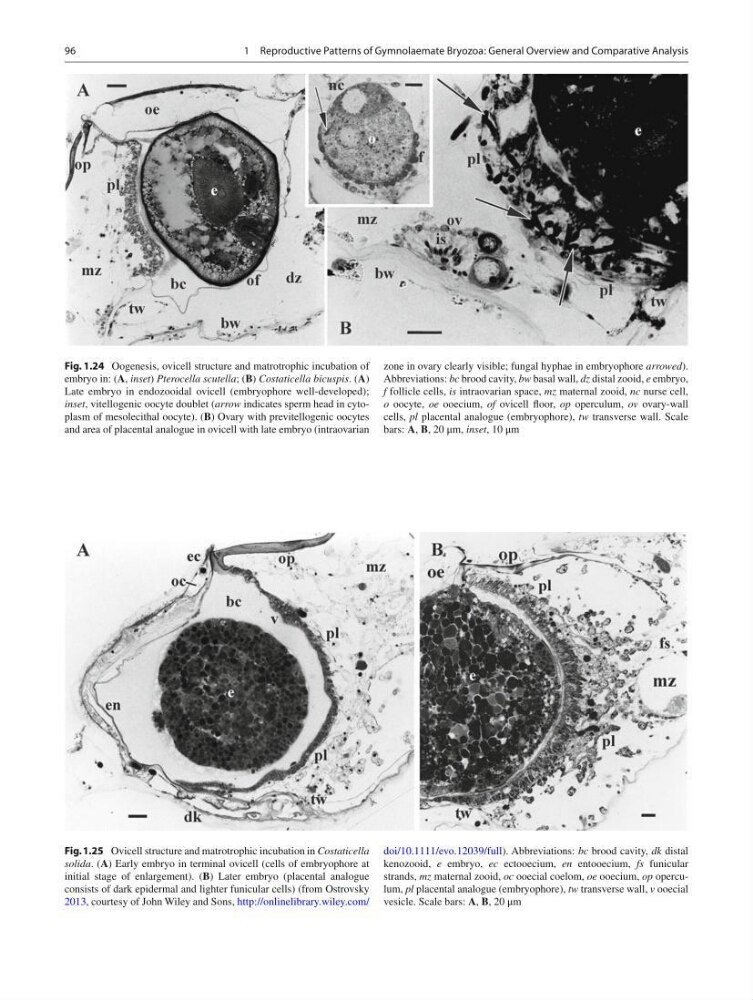
Fig. 1.24 Oogenesis, ovicell structure and matrotrophic incubation of
embryo in: ( A , inset ) Pterocella scutella ; ( B ) Costaticella bicuspis . ( A )
Late embryo in endozooidal ovicell (embryophore well-developed);
inset , vitellogenic oocyte doublet ( arrow indicates sperm head in cytoplasm of mesolecithal oocyte). ( B ) Ovary with previtellogenic oocytes
and area of placental analogue in ovicell with late embryo (intraovarian
zone in ovary clearly visible; fungal hyphae in embryophore arrowed ).
Abbreviations: bc brood cavity, bw basal wall, dz distal zooid, е embryo,
f follicle cells, is intraovarian space, mz maternal zooid, nc nurse cell,
о oocyte, ое ooecium, of ovicell fl oor, op operculum, ov ovary-wall
cells, pl placental analogue (embryophore), tw transverse wall. Scale
bars: A , B , 20 μm, inset , 10 μm
Fig. 1.25 Ovicell structure and matrotrophic incubation in Costaticella
solida . ( A ) Early embryo in terminal ovicell (cells of embryophore at
initial stage of enlargement). ( B ) Later embryo (placental analogue
consists of dark epidermal and lighter funicular cells) (from Ostrovsky
2013 , courtesy of John Wiley and Sons, http://onlinelibrary.wiley.com/
doi/10.1111/evo.12039/full ). Abbreviations: bc brood cavity, dk distal
kenozooid, е embryo, ec ectooecium, en entooecium, fs funicular
strands, mz maternal zooid, ос ooecial coelom, ое ooecium, op operculum, pl placental analogue (embryophore), tw transverse wall, v ooecial
vesicle. Scale bars: A , B , 20 μm
1 Reproductive Patterns of Gymnolaemate Bryozoa: General Overview and Comparative Analysis
Fig. 1.24 Oogenesis, ovicell structure and matrotrophic incubation of
embryo in: ( A , inset ) Pterocella scutella ; ( B ) Costaticella bicuspis . ( A )
Late embryo in endozooidal ovicell (embryophore well-developed);
inset , vitellogenic oocyte doublet ( arrow indicates sperm head in cytoplasm of mesolecithal oocyte). ( B ) Ovary with previtellogenic oocytes
and area of placental analogue in ovicell with late embryo (intraovarian
zone in ovary clearly visible; fungal hyphae in embryophore arrowed ).
Abbreviations: bc brood cavity, bw basal wall, dz distal zooid, е embryo,
f follicle cells, is intraovarian space, mz maternal zooid, nc nurse cell,
о oocyte, ое ooecium, of ovicell fl oor, op operculum, ov ovary-wall
cells, pl placental analogue (embryophore), tw transverse wall. Scale
bars: A , B , 20 μm, inset , 10 μm
Fig. 1.25 Ovicell structure and matrotrophic incubation in Costaticella
solida . ( A ) Early embryo in terminal ovicell (cells of embryophore at
initial stage of enlargement). ( B ) Later embryo (placental analogue
consists of dark epidermal and lighter funicular cells) (from Ostrovsky
2013 , courtesy of John Wiley and Sons, http://onlinelibrary.wiley.com/
doi/10.1111/evo.12039/full ). Abbreviations: bc brood cavity, dk distal
kenozooid, е embryo, ec ectooecium, en entooecium, fs funicular
strands, mz maternal zooid, ос ooecial coelom, ое ooecium, op operculum, pl placental analogue (embryophore), tw transverse wall, v ooecial
vesicle. Scale bars: A , B , 20 μm
1 Reproductive Patterns of Gymnolaemate Bryozoa: General Overview and Comparative Analysis