

87
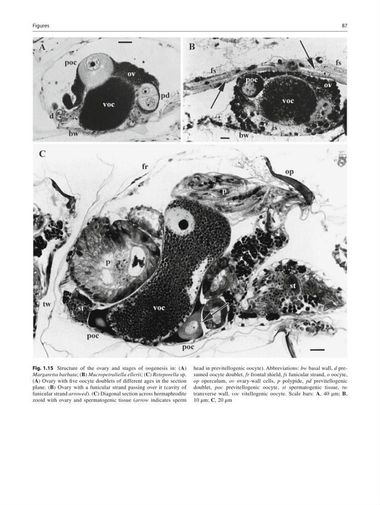
Fig. 1.15 Structure of the ovary and stages of oogenesis in: ( A )
Margaretta barbata ; ( B ) Mucropetraliella ellerii ; ( C ) Reteporella sp.
( A ) Ovary with fi ve oocyte doublets of different ages in the section
plane. ( B ) Ovary with a funicular strand passing over it (cavity of
funicular strand arrowed ). ( C ) Diagonal section across hermaphrodite
zooid with ovary and spermatogenic tissue ( arrow indicates sperm
head in previtellogenic oocyte). Abbreviations: bw basal wall, d presumed oocyte doublet, fr frontal shield, fs funicular strand, o oocyte,
op operculum, ov ovary-wall cells, p polypide, рd previtellogenic
doublet, poc previtellogenic oocyte, st spermatogenic tissue, tw
transverse wall, voc vitellogenic oocyte. Scale bars: A , 40 μm; B ,
10 μm; C , 20 μm
Figures
Fig. 1.15 Structure of the ovary and stages of oogenesis in: ( A )
Margaretta barbata ; ( B ) Mucropetraliella ellerii ; ( C ) Reteporella sp.
( A ) Ovary with fi ve oocyte doublets of different ages in the section
plane. ( B ) Ovary with a funicular strand passing over it (cavity of
funicular strand arrowed ). ( C ) Diagonal section across hermaphrodite
zooid with ovary and spermatogenic tissue ( arrow indicates sperm
head in previtellogenic oocyte). Abbreviations: bw basal wall, d presumed oocyte doublet, fr frontal shield, fs funicular strand, o oocyte,
op operculum, ov ovary-wall cells, p polypide, рd previtellogenic
doublet, poc previtellogenic oocyte, st spermatogenic tissue, tw
transverse wall, voc vitellogenic oocyte. Scale bars: A , 40 μm; B ,
10 μm; C , 20 μm
Figures