

85
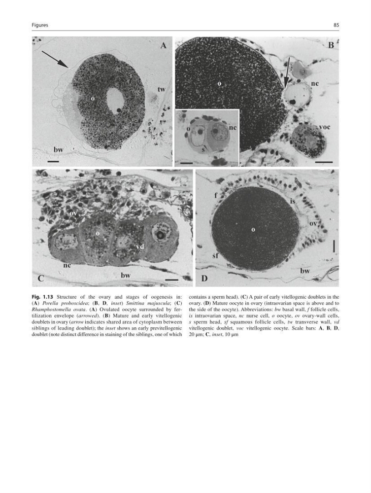
Fig. 1.13 Structure of the ovary and stages of oogenesis in:
( A ) Porella proboscidea ; ( B , D , inset ) Smittina majuscula ; ( C )
Rhamphostomella ovata . ( A ) Ovulated oocyte surrounded by fertilization envelope ( arrowed ). ( B ) Mature and early vitellogenic
doublets in ovary ( arrow indicates shared area of cytoplasm between
siblings of leading doublet); the inset shows an early previtellogenic
doublet (note distinct difference in staining of the siblings, one of which
contains a sperm head). ( C ) A pair of early vitellogenic doublets in the
ovary. ( D ) Mature oocyte in ovary (intraovarian space is above and to
the side of the oocyte). Abbreviations: bw basal wall, f follicle cells,
is intraovarian space, nc nurse cell, o oocyte, ov ovary- wall cells,
s sperm head, sf squamous follicle cells, tw transverse wall, vd
vitellogenic doublet, voc vitellogenic oocyte. Scale bars: A , B , D ,
20 μm; C , inset , 10 μm
Figures
Fig. 1.13 Structure of the ovary and stages of oogenesis in:
( A ) Porella proboscidea ; ( B , D , inset ) Smittina majuscula ; ( C )
Rhamphostomella ovata . ( A ) Ovulated oocyte surrounded by fertilization envelope ( arrowed ). ( B ) Mature and early vitellogenic
doublets in ovary ( arrow indicates shared area of cytoplasm between
siblings of leading doublet); the inset shows an early previtellogenic
doublet (note distinct difference in staining of the siblings, one of which
contains a sperm head). ( C ) A pair of early vitellogenic doublets in the
ovary. ( D ) Mature oocyte in ovary (intraovarian space is above and to
the side of the oocyte). Abbreviations: bw basal wall, f follicle cells,
is intraovarian space, nc nurse cell, o oocyte, ov ovary- wall cells,
s sperm head, sf squamous follicle cells, tw transverse wall, vd
vitellogenic doublet, voc vitellogenic oocyte. Scale bars: A , B , D ,
20 μm; C , inset , 10 μm
Figures