

79
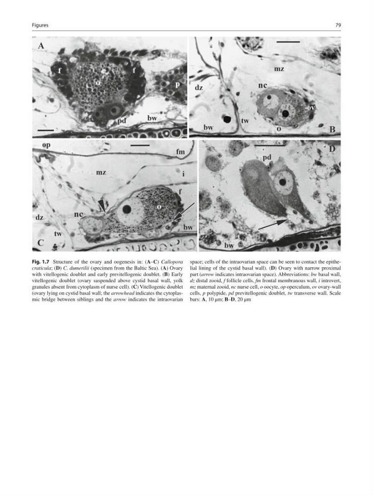
Fig. 1.7 Structure of the ovary and oogenesis in: ( A – C ) Callopora
craticula ; ( D ) C . dumerilii (specimen from the Baltic Sea). ( A ) Ovary
with vitellogenic doublet and early previtellogenic doublet. ( B ) Early
vitellogenic doublet (ovary suspended above cystid basal wall, yolk
granules absent from cytoplasm of nurse cell). ( C ) Vitellogenic doublet
(ovary lying on cystid basal wall; the arrowhead indicates the cytoplasmic bridge between siblings and the arrow indicates the intraovarian
space; cells of the intraovarian space can be seen to contact the epithelial lining of the cystid basal wall). ( D ) Ovary with narrow proximal
part ( arrow indicates intraovarian space). Abbreviations: bw basal wall,
dz distal zooid, f follicle cells, fm frontal membranous wall, i introvert,
mz maternal zooid, nc nurse cell, o oocyte, ор operculum, ov ovary-wall
cells, р polypide, pd previtellogenic doublet, tw transverse wall. Scale
bars: A , 10 μm; B – D , 20 μm
Figures
Fig. 1.7 Structure of the ovary and oogenesis in: ( A – C ) Callopora
craticula ; ( D ) C . dumerilii (specimen from the Baltic Sea). ( A ) Ovary
with vitellogenic doublet and early previtellogenic doublet. ( B ) Early
vitellogenic doublet (ovary suspended above cystid basal wall, yolk
granules absent from cytoplasm of nurse cell). ( C ) Vitellogenic doublet
(ovary lying on cystid basal wall; the arrowhead indicates the cytoplasmic bridge between siblings and the arrow indicates the intraovarian
space; cells of the intraovarian space can be seen to contact the epithelial lining of the cystid basal wall). ( D ) Ovary with narrow proximal
part ( arrow indicates intraovarian space). Abbreviations: bw basal wall,
dz distal zooid, f follicle cells, fm frontal membranous wall, i introvert,
mz maternal zooid, nc nurse cell, o oocyte, ор operculum, ov ovary-wall
cells, р polypide, pd previtellogenic doublet, tw transverse wall. Scale
bars: A , 10 μm; B – D , 20 μm
Figures