

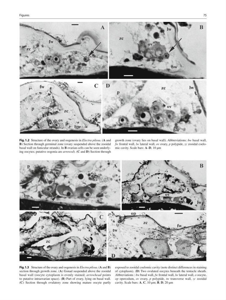
Fig. 1.2 Structure of the ovary and oogenesis in Electra pilosa . ( A and
B ) Section through germinal zone (ovary suspended above the zooidal
basal wall on funicular strands). In B ovarian cells can be seen underlying oocytes; putative oogonia are arrowed ). ( C and D ) Section through
growth zone (ovary lies on basal wall). Abbreviations: bw basal wall,
fw frontal wall, lw lateral wall, ov ovary, p polypide, zc zooidal coelomic cavity. Scale bars: A – D , 10 μm
Fig. 1.3 Structure of the ovary and oogenesis in Electra pilosa . ( A and B )
section through growth zone. ( A ) Gonad suspended above the zooidal
basal wall (oocyte cytoplasm is evenly stained; arrowhead points
to putative intraovarian space). ( B ) Part of ovary, lying on basal wall.
( C ) Section through ovulatory zone showing mature oocyte partly
exposed to zooidal coelomic cavity (note distinct differences in staining
of cytoplasm). ( D ) Two ovulated oocytes beneath the tentacle sheath.
Abbreviations : bw basal wall, fw frontal wall, lw lateral wall, о oocyte,
op operculum, ov ovary, p polypide, tw transverse wall, zc zooidal
cavity. Scale bars: A , C , 10 μm; B , D , 20 μm
75
Figures
B ) Section through germinal zone (ovary suspended above the zooidal
basal wall on funicular strands). In B ovarian cells can be seen underlying oocytes; putative oogonia are arrowed ). ( C and D ) Section through
growth zone (ovary lies on basal wall). Abbreviations: bw basal wall,
fw frontal wall, lw lateral wall, ov ovary, p polypide, zc zooidal coelomic cavity. Scale bars: A – D , 10 μm
Fig. 1.3 Structure of the ovary and oogenesis in Electra pilosa . ( A and B )
section through growth zone. ( A ) Gonad suspended above the zooidal
basal wall (oocyte cytoplasm is evenly stained; arrowhead points
to putative intraovarian space). ( B ) Part of ovary, lying on basal wall.
( C ) Section through ovulatory zone showing mature oocyte partly
exposed to zooidal coelomic cavity (note distinct differences in staining
of cytoplasm). ( D ) Two ovulated oocytes beneath the tentacle sheath.
Abbreviations : bw basal wall, fw frontal wall, lw lateral wall, о oocyte,
op operculum, ov ovary, p polypide, tw transverse wall, zc zooidal
cavity. Scale bars: A , C , 10 μm; B , D , 20 μm
75
Figures