

67
interpreted from the observations of Hincks ( 1851 ) that the
ITO could have the additional function of sperm release.
They speculated that this could enable expulsion of the
remaining sperm at the end of the reproductive period. Later,
Temkin ( 1994 ) suggested that squeezing of the inseminated
oocyte through the ITO may trigger activation of the egg by
either (1) physical stress or (2) chemical stimulation. It was
also shown that both the passage of eggs and the entering of
sperm via the ITO is regulated by a terminal sphincter muscle in M . membranacea (Temkin 1994 ).
The presence of an ITO is strongly correlated with broadcasting reproductive pattern I, involving production of
numerous small yolk-poor eggs that develop into long-lived
planktotrophic larvae (see Sect. 1.2.4 ). This pattern of reproduction is considered to be ancestral by most scholars, and it
is also rather rare (but probably underreported) among
Bryozoa. Within the Gymnolaemata it is typical of the earliest cheilostome group, Malacostegina, with primitive skeletal organization, and also broadcasting ctenostomes, being
absent from the Stenolaemata and Phylactolaemata (Ryland
1970 , 1976 ; Reed 1991 ). A few gymnolaemate brooders also
have an ITO and a few broadcasters have a SNP, but these are
exceptions (discussed below).
1.3.9.1 Competing Hypotheses on the Origin
and the Function of the Intertentacular
Organ
According to the Silén’s ( 1945 ) hypothesis, the ITO and SNP
evolved by transformation of two tentacles through their
fusion and “shortening”: an intertentacular organ might have
evolved at the expense of those tentacles with terminal
(coelomo)pores, becoming reduced at a later stage to a simple
pore. Ostrovsky and Porter ( 2011 ) proposed an alternative
hypothesis, in which the ITO evolved from a female gonopore as an extension developing from the fusion of the basal
parts of two dorso-medial tentacles. This is in accordance
with the development of the ITO as described by Hageman
( 1981 ) and Reed ( 1991 ) who called the ITO an extension of
the female gonopore.
Silén ( 1944 ) speculated that in Phylactolaemata and primitive brooding Gymnolaemata (such as Labiostomella gisleni ),
the ovulated egg never leaves the zooidal cavity and enters an
“embryonary” or “embryo sac” formed on the internal surface of the maternal zooid body wall. Upon maturation, the
larva is released from this brood chamber either by rupture of
the body wall or through the zooidal orifi ce after polypide
degeneration. In this scenario there was no requirement for a
female “birth” pore in ancestral bryozoans, although there is
a need for a pore that allows entry of allosperm. The simultaneous presence of the SNP and “embryonary” in ctenostomes
of the genus Nolella led Silén ( 1945 ) to develop the idea that
the ITO and SNP initially evolved not for the spawning of
eggs but for the entry of sperm, secondarily acquiring a
spawning function in descendants. According to Cori ( 1941 ),
the ITO is formed by the fusion of two neighbouring tentacles. This led Silén ( 1945 , p. 25) to suggest that the ITO and
SNP were homologous with tentacles and comparable to terminal tentacle pores, and that sperm might enter a zooidal
coelom via the tentacle pores. Notably, Cori ( 1941 ) recorded
spermatozoids in the coelomic lumen of the tentacles in the
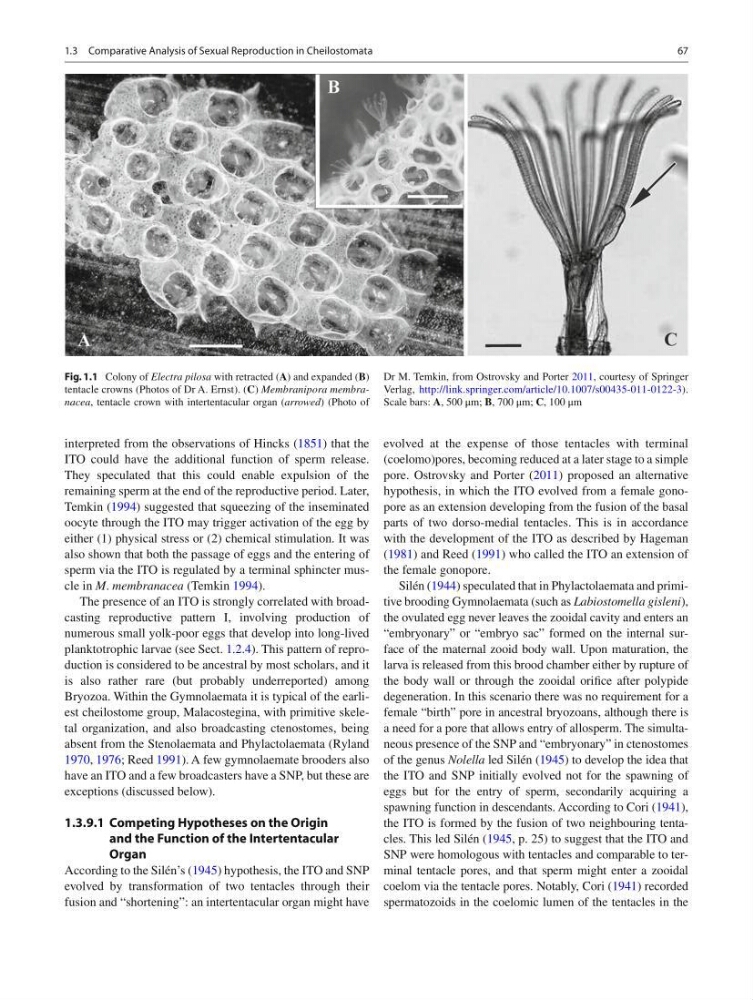
Fig. 1.1 Colony of Electra pilosa with retracted ( A ) and expanded ( B )
tentacle crowns (Photos of Dr A. Ernst). ( C ) Membranipora membranacea , tentacle crown with intertentacular organ ( arrowed ) (Photo of
Dr M. Temkin, from Ostrovsky and Porter 2011 , courtesy of Springer
Verlag, http://link.springer.com/article/10.1007/s00435-011-0122-3 ).
Scale bars: A , 500 μm; B , 700 μm; C , 100 μm
1.3 Comparative Analysis of Sexual Reproduction in Cheilostomata
interpreted from the observations of Hincks ( 1851 ) that the
ITO could have the additional function of sperm release.
They speculated that this could enable expulsion of the
remaining sperm at the end of the reproductive period. Later,
Temkin ( 1994 ) suggested that squeezing of the inseminated
oocyte through the ITO may trigger activation of the egg by
either (1) physical stress or (2) chemical stimulation. It was
also shown that both the passage of eggs and the entering of
sperm via the ITO is regulated by a terminal sphincter muscle in M . membranacea (Temkin 1994 ).
The presence of an ITO is strongly correlated with broadcasting reproductive pattern I, involving production of
numerous small yolk-poor eggs that develop into long-lived
planktotrophic larvae (see Sect. 1.2.4 ). This pattern of reproduction is considered to be ancestral by most scholars, and it
is also rather rare (but probably underreported) among
Bryozoa. Within the Gymnolaemata it is typical of the earliest cheilostome group, Malacostegina, with primitive skeletal organization, and also broadcasting ctenostomes, being
absent from the Stenolaemata and Phylactolaemata (Ryland
1970 , 1976 ; Reed 1991 ). A few gymnolaemate brooders also
have an ITO and a few broadcasters have a SNP, but these are
exceptions (discussed below).
1.3.9.1 Competing Hypotheses on the Origin
and the Function of the Intertentacular
Organ
According to the Silén’s ( 1945 ) hypothesis, the ITO and SNP
evolved by transformation of two tentacles through their
fusion and “shortening”: an intertentacular organ might have
evolved at the expense of those tentacles with terminal
(coelomo)pores, becoming reduced at a later stage to a simple
pore. Ostrovsky and Porter ( 2011 ) proposed an alternative
hypothesis, in which the ITO evolved from a female gonopore as an extension developing from the fusion of the basal
parts of two dorso-medial tentacles. This is in accordance
with the development of the ITO as described by Hageman
( 1981 ) and Reed ( 1991 ) who called the ITO an extension of
the female gonopore.
Silén ( 1944 ) speculated that in Phylactolaemata and primitive brooding Gymnolaemata (such as Labiostomella gisleni ),
the ovulated egg never leaves the zooidal cavity and enters an
“embryonary” or “embryo sac” formed on the internal surface of the maternal zooid body wall. Upon maturation, the
larva is released from this brood chamber either by rupture of
the body wall or through the zooidal orifi ce after polypide
degeneration. In this scenario there was no requirement for a
female “birth” pore in ancestral bryozoans, although there is
a need for a pore that allows entry of allosperm. The simultaneous presence of the SNP and “embryonary” in ctenostomes
of the genus Nolella led Silén ( 1945 ) to develop the idea that
the ITO and SNP initially evolved not for the spawning of
eggs but for the entry of sperm, secondarily acquiring a
spawning function in descendants. According to Cori ( 1941 ),
the ITO is formed by the fusion of two neighbouring tentacles. This led Silén ( 1945 , p. 25) to suggest that the ITO and
SNP were homologous with tentacles and comparable to terminal tentacle pores, and that sperm might enter a zooidal
coelom via the tentacle pores. Notably, Cori ( 1941 ) recorded
spermatozoids in the coelomic lumen of the tentacles in the
Fig. 1.1 Colony of Electra pilosa with retracted ( A ) and expanded ( B )
tentacle crowns (Photos of Dr A. Ernst). ( C ) Membranipora membranacea , tentacle crown with intertentacular organ ( arrowed ) (Photo of
Dr M. Temkin, from Ostrovsky and Porter 2011 , courtesy of Springer
Verlag, http://link.springer.com/article/10.1007/s00435-011-0122-3 ).
Scale bars: A , 500 μm; B , 700 μm; C , 100 μm
1.3 Comparative Analysis of Sexual Reproduction in Cheilostomata