

~_~~--..s:· 1 and chI
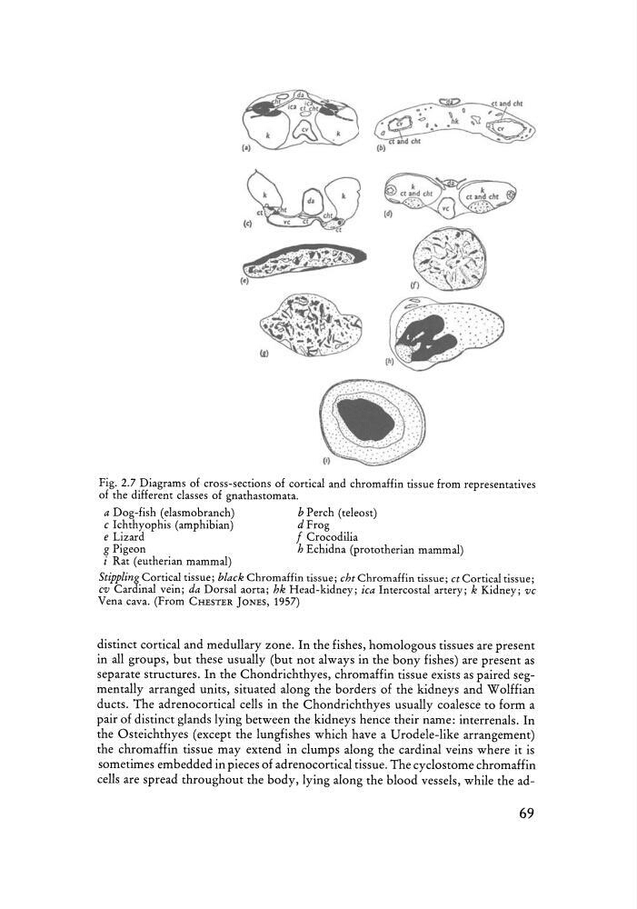
Fig. 2.7 Diagrams of cros s-sections of cortical and chromaffin tissue from repre sentatives
of the different classes of gnathasto mata.
a Dog-fish (elasrnobranch)
b Perch (teleo st)
c Ichthyophis (amphibian)
d Fro g
e Lizard
f Crocodilia
g Pigeon
h Echidna (pro tot herian mammal)
i Rat (eutherian mammal)
Stippling Cortical tissue; black Chromaffin tissue; cht Ch rom affin tissue; ct Cortical tissue;
cu Cardinal vein; da Dorsal aorta; hk Head-kidne y ; ica Intercostal artery ; k Kidney; vc
Vena cava. (From CHESTER JONES, 1957)
distinct cortical and medullary zone. In the fishes, homologous tissues are present
in all groups, but these usually (but not always in the bony fishes) are present as
separate structures. In the Chondrichthyes, chromaffin tissue exists as paired segmentally arranged units, situ ated along the borders of the kidneys and W olffian
ducts. The adrenocortical cells in the Chondrichthyes usuall y coalesce to form a
pair of distinct glands lyin g between the kidneys henc e th eir name : interrenals. In
the Osteichthyes (except the lungfishes which have a Urodele-like arrangement)
the chromaffin tissu e may extend in clumps along the cardinal veins where it is
sometimes embedded in pieces of adrenocortical tissue. The cyclostome chromaffin
cells are spread throughout the bod y, lyin g along the blood vessels, while the ad69
Fig. 2.7 Diagrams of cros s-sections of cortical and chromaffin tissue from repre sentatives
of the different classes of gnathasto mata.
a Dog-fish (elasrnobranch)
b Perch (teleo st)
c Ichthyophis (amphibian)
d Fro g
e Lizard
f Crocodilia
g Pigeon
h Echidna (pro tot herian mammal)
i Rat (eutherian mammal)
Stippling Cortical tissue; black Chromaffin tissue; cht Ch rom affin tissue; ct Cortical tissue;
cu Cardinal vein; da Dorsal aorta; hk Head-kidne y ; ica Intercostal artery ; k Kidney; vc
Vena cava. (From CHESTER JONES, 1957)
distinct cortical and medullary zone. In the fishes, homologous tissues are present
in all groups, but these usually (but not always in the bony fishes) are present as
separate structures. In the Chondrichthyes, chromaffin tissue exists as paired segmentally arranged units, situ ated along the borders of the kidneys and W olffian
ducts. The adrenocortical cells in the Chondrichthyes usuall y coalesce to form a
pair of distinct glands lyin g between the kidneys henc e th eir name : interrenals. In
the Osteichthyes (except the lungfishes which have a Urodele-like arrangement)
the chromaffin tissu e may extend in clumps along the cardinal veins where it is
sometimes embedded in pieces of adrenocortical tissue. The cyclostome chromaffin
cells are spread throughout the bod y, lyin g along the blood vessels, while the ad69