

that act directly on effector tissues, and it also serves as the major link connecting
the nervous and endocrine systems. Information concerning conditions in the external and internal environments is transmitted from the neural elements of the
brain to the pituitary, where it is translated into relevant endocrine language which
is then dispatched to the appropriate effector tissues. The external neural information comes from such things as light, temperature, the presence of the opposite
sex and otherwise stressful or potenially harmful situations. Internal information
about the general homeostatic state of the animal, such as water, solute and hormone levels in th e body fluids, is also transmitted through the brain.
a) Morphology
Phyletically the pituitary gland can first be distinguished in the cyclostome fishes
but is absent in protochordates like Amphioxus. The gland arises from two types
of tissue; ectoderm from the anterior region of the head (hypophysis) and neural
tissue (infundibulum) arising as a downgrowth from the forebrain . The glandular
parts of these tissues remain in a juxtaposition with the hypothalamic region of
the brain just behind the optic chiasma. In the Agnatha the different regions of
the pituitary are separate, while in other vertebrates they are usually more tightly
associated, but still retain their separate endocrine integrity and connections with
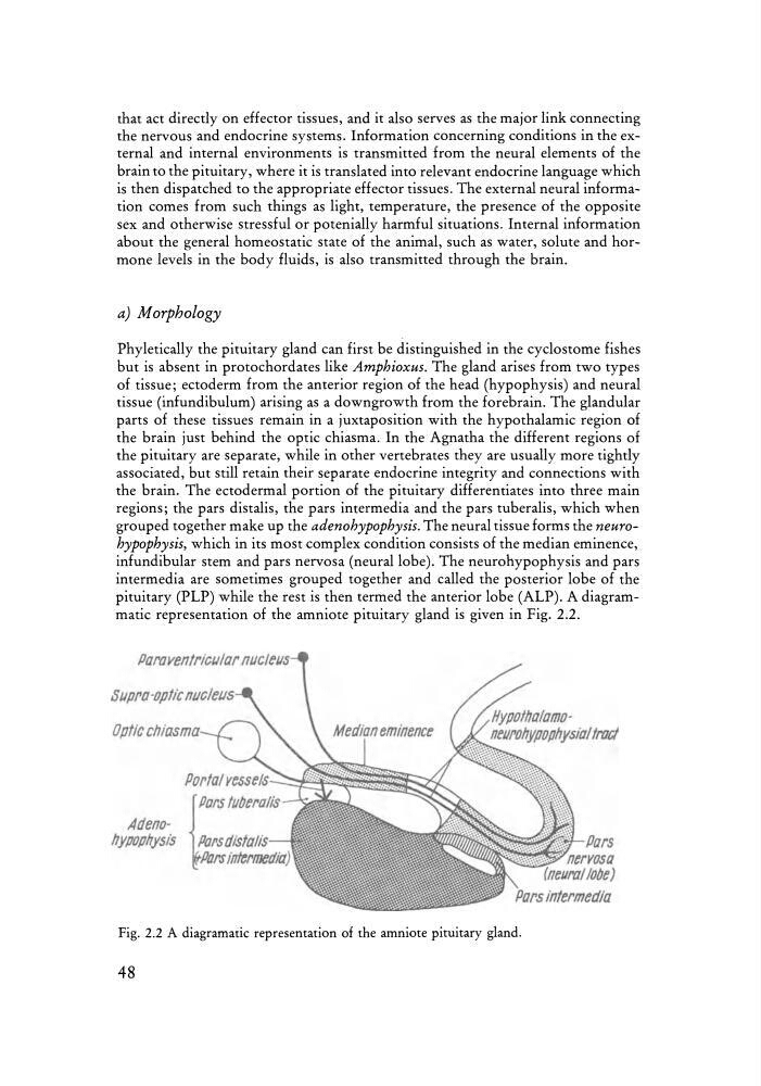
the brain. The ectodermal portion of the pituitary differentiates into three main
regions; the pars distalis, the pars intermedia and the pars tuberalis, which when
grouped together make up the adenohypophysis. The neural tissue forms the neurohypophysis, which in its most complex condition consists of the median eminence,
infundibular stem and pars nervosa (neural lobe). The neurohypophysis and pars
intermedia are sometimes grouped together and called the posterior lobe of the
pituitary (PLP) while the rest is then termed the anterior lobe (ALP). A diagrammatic representation of the amniote pituitary gland is given in Fig. 2.2.
aaravenmcutar nucleus
Optic chiasma
Portal Yesse/.s~-::f.~~~~~S~
{
Pars tllbera/is
Adenohypophysis Parsdista/is
Parsintermedia)
Fig. 2.2 A diagramatic representation of the amniote pituitary gland.
48
the nervous and endocrine systems. Information concerning conditions in the external and internal environments is transmitted from the neural elements of the
brain to the pituitary, where it is translated into relevant endocrine language which
is then dispatched to the appropriate effector tissues. The external neural information comes from such things as light, temperature, the presence of the opposite
sex and otherwise stressful or potenially harmful situations. Internal information
about the general homeostatic state of the animal, such as water, solute and hormone levels in th e body fluids, is also transmitted through the brain.
a) Morphology
Phyletically the pituitary gland can first be distinguished in the cyclostome fishes
but is absent in protochordates like Amphioxus. The gland arises from two types
of tissue; ectoderm from the anterior region of the head (hypophysis) and neural
tissue (infundibulum) arising as a downgrowth from the forebrain . The glandular
parts of these tissues remain in a juxtaposition with the hypothalamic region of
the brain just behind the optic chiasma. In the Agnatha the different regions of
the pituitary are separate, while in other vertebrates they are usually more tightly
associated, but still retain their separate endocrine integrity and connections with
the brain. The ectodermal portion of the pituitary differentiates into three main
regions; the pars distalis, the pars intermedia and the pars tuberalis, which when
grouped together make up the adenohypophysis. The neural tissue forms the neurohypophysis, which in its most complex condition consists of the median eminence,
infundibular stem and pars nervosa (neural lobe). The neurohypophysis and pars
intermedia are sometimes grouped together and called the posterior lobe of the
pituitary (PLP) while the rest is then termed the anterior lobe (ALP). A diagrammatic representation of the amniote pituitary gland is given in Fig. 2.2.
aaravenmcutar nucleus
Optic chiasma
Portal Yesse/.s~-::f.~~~~~S~
{
Pars tllbera/is
Adenohypophysis Parsdista/is
Parsintermedia)
Fig. 2.2 A diagramatic representation of the amniote pituitary gland.
48