

82
3 The Structural Variety and Metabolism of Proteins
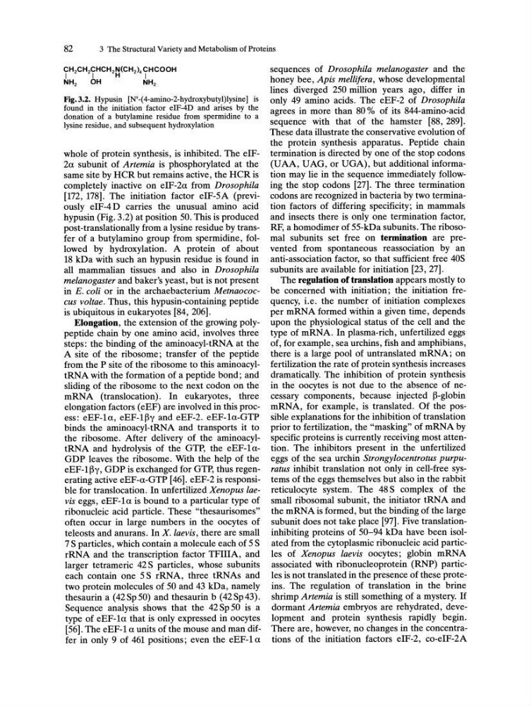
Fig.3.2. Hypusin [N
E
_( 4-amino-2-hydroxybutyl)lysine] is
found in the initiation factor eIF-4D and arises by the
donation of a butylamine residue from spermidine to a
lysine residue, and subsequent hydroxylation
whole of protein synthesis, is inhibited. The elF2a subunit of Artemia is phosphorylated at the
same site by HCR but remains active, the HCR is
completely inactive on elF-2a from Drosophila
[172, 178]. The initiation factor eIF-5A (previously e1F-4 D carries the unusual amino acid
hypusin (Fig. 3.2) at position 50. This is produced
post-translationally from a lysine residue by transfer of a butylamino group from spermidine, followed by hydroxylation. A protein of about
18 kDa with such an hypusin residue is found in
all mammalian tissues and also in Drosophila
melanogaster and baker's yeast, but is not present
in E. coli or in the archaebacterium Metnaococcus voltae. Thus, this hypusin-containing peptide
is ubiquitous in eukaryotes [84, 206].
Elongation, the extension of the growing polypeptide chain by one amino acid, involves three
steps: the binding of the aminoacyl-tRNA at the
A site of the ribosome; transfer of the peptide
from the P site of the ribosome to this aminoacyltRNA with the formation of a peptide bond; and
sliding of the ribosome to the next codon on the
mRNA (translocation). In eukaryotes, three
elongation factors (eEF) are involved in this process: eEF-la, eEF-l~y and eEF-2. eEF-la-GTP
binds the aminoacyl-tRNA and transports it to
the ribosome. After delivery of the aminoacyltRNA and hydrolysis of the GTP, the eEF-laGDP leaves the ribosome. With the help of the
eEF-l~y, GDP is exchanged for GTP, thus regenerating active eEF-a-GTP [46]. eEF-2 is responsible for translocation. In unfertilized Xenopus laevis eggs, eEF-la is bound to a particular type of
ribonucleic acid particle. These "thesaurisomes"
often occur in large numbers in the oocytes of
teleosts and anurans. In X. laevis, there are small
7 S particles, which contain a molecule each of 5 S
rRNA and the transcription factor TFIIIA, and
larger tetrameric 42 S particles, whose subunits
each contain one 5 S rRNA, three tRNAs and
two protein molecules of 50 and 43 kDa, namely
thesaurin a (42Sp50) and thesaurin b (42Sp43).
Sequence analysis shows that the 42 Sp 50 is a
type of eEF-la that is only expressed in oocytes
[56]. The eEF-l a units of the mouse and man differ in only 9 of 461 positions; even the eEF-l a
sequences of Drosophila melanogaster and the
honey bee, Apis melli/era, whose developmental
lines diverged 250 million years ago, differ in
only 49 amino acids. The eEF-2 of Drosophila
agrees in more than 80 % of its 844-amino-acid
sequence with that of the hamster [88,289].
These data illustrate the conservative evolution of
the protein synthesis apparatus. Peptide chain
termination is directed by one of the stop codons
(UAA, UAG, or UGA), but additional information may lie in the sequence immediately following the stop codons [27]. The three termination
codons are recognized in bacteria by two termination factors of differing specificity; in mammals
and insects there is only one termination factor,
RF, a homodimer of 55-kDa subunits. The ribosomal subunits set free on termination are prevented from spontaneous reassociation by an
anti-association factor, so that sufficient free 40S
subunits are available for initiation [23, 27].
The regulation of translation appears mostly to
be concerned with initiation; the initiation frequency, i.e. the number of initiation complexes
per mRNA formed within a given time, depends
upon the physiological status of the cell and the
type of mRNA. In plasma-rich, unfertilized eggs
of, for example, sea urchins, fish and amphibians,
there is a large pool of untranslated mRNA; on
fertilization the rate of protein synthesis increases
dramatically. The inhibition of protein synthesis
in the oocytes is not due to the absence of necessary components, because injected ~-globin
mRNA, for example, is translated. Of the possible explanations for the inhibition of translation
prior to fertilization, the "masking" of mRNA by
specific proteins is currently receiving most attention. The inhibitors present in the unfertilized
eggs of the sea urchin Strongylocentrotus purpuratus inhibit translation not only in cell-free systems of the eggs themselves but also in the rabbit
reticulocyte system. The 48 S complex of the
small ribosomal subunit, the initiator tRNA and
the mRNA is formed, but the binding of the large
subunit does not take place [97]. Five translationinhibiting proteins of 50-94 kDa have been isolated from the cytoplasmic ribonucleic acid particles of Xenopus laevis oocytes; globin mRNA
associated with ribonucleoprotein (RNP) particles is not translated in the presence of these proteins. The regulation of translation in the brine
shrimp Artemia is still something of a mystery. If
dormant Artemia embryos are rehydrated, development and protein synthesis rapidly begin.
There are, however, no changes in the concentrations of the initiation factors eIF-2, co-eIF-2A
3 The Structural Variety and Metabolism of Proteins
Fig.3.2. Hypusin [N
E
_( 4-amino-2-hydroxybutyl)lysine] is
found in the initiation factor eIF-4D and arises by the
donation of a butylamine residue from spermidine to a
lysine residue, and subsequent hydroxylation
whole of protein synthesis, is inhibited. The elF2a subunit of Artemia is phosphorylated at the
same site by HCR but remains active, the HCR is
completely inactive on elF-2a from Drosophila
[172, 178]. The initiation factor eIF-5A (previously e1F-4 D carries the unusual amino acid
hypusin (Fig. 3.2) at position 50. This is produced
post-translationally from a lysine residue by transfer of a butylamino group from spermidine, followed by hydroxylation. A protein of about
18 kDa with such an hypusin residue is found in
all mammalian tissues and also in Drosophila
melanogaster and baker's yeast, but is not present
in E. coli or in the archaebacterium Metnaococcus voltae. Thus, this hypusin-containing peptide
is ubiquitous in eukaryotes [84, 206].
Elongation, the extension of the growing polypeptide chain by one amino acid, involves three
steps: the binding of the aminoacyl-tRNA at the
A site of the ribosome; transfer of the peptide
from the P site of the ribosome to this aminoacyltRNA with the formation of a peptide bond; and
sliding of the ribosome to the next codon on the
mRNA (translocation). In eukaryotes, three
elongation factors (eEF) are involved in this process: eEF-la, eEF-l~y and eEF-2. eEF-la-GTP
binds the aminoacyl-tRNA and transports it to
the ribosome. After delivery of the aminoacyltRNA and hydrolysis of the GTP, the eEF-laGDP leaves the ribosome. With the help of the
eEF-l~y, GDP is exchanged for GTP, thus regenerating active eEF-a-GTP [46]. eEF-2 is responsible for translocation. In unfertilized Xenopus laevis eggs, eEF-la is bound to a particular type of
ribonucleic acid particle. These "thesaurisomes"
often occur in large numbers in the oocytes of
teleosts and anurans. In X. laevis, there are small
7 S particles, which contain a molecule each of 5 S
rRNA and the transcription factor TFIIIA, and
larger tetrameric 42 S particles, whose subunits
each contain one 5 S rRNA, three tRNAs and
two protein molecules of 50 and 43 kDa, namely
thesaurin a (42Sp50) and thesaurin b (42Sp43).
Sequence analysis shows that the 42 Sp 50 is a
type of eEF-la that is only expressed in oocytes
[56]. The eEF-l a units of the mouse and man differ in only 9 of 461 positions; even the eEF-l a
sequences of Drosophila melanogaster and the
honey bee, Apis melli/era, whose developmental
lines diverged 250 million years ago, differ in
only 49 amino acids. The eEF-2 of Drosophila
agrees in more than 80 % of its 844-amino-acid
sequence with that of the hamster [88,289].
These data illustrate the conservative evolution of
the protein synthesis apparatus. Peptide chain
termination is directed by one of the stop codons
(UAA, UAG, or UGA), but additional information may lie in the sequence immediately following the stop codons [27]. The three termination
codons are recognized in bacteria by two termination factors of differing specificity; in mammals
and insects there is only one termination factor,
RF, a homodimer of 55-kDa subunits. The ribosomal subunits set free on termination are prevented from spontaneous reassociation by an
anti-association factor, so that sufficient free 40S
subunits are available for initiation [23, 27].
The regulation of translation appears mostly to
be concerned with initiation; the initiation frequency, i.e. the number of initiation complexes
per mRNA formed within a given time, depends
upon the physiological status of the cell and the
type of mRNA. In plasma-rich, unfertilized eggs
of, for example, sea urchins, fish and amphibians,
there is a large pool of untranslated mRNA; on
fertilization the rate of protein synthesis increases
dramatically. The inhibition of protein synthesis
in the oocytes is not due to the absence of necessary components, because injected ~-globin
mRNA, for example, is translated. Of the possible explanations for the inhibition of translation
prior to fertilization, the "masking" of mRNA by
specific proteins is currently receiving most attention. The inhibitors present in the unfertilized
eggs of the sea urchin Strongylocentrotus purpuratus inhibit translation not only in cell-free systems of the eggs themselves but also in the rabbit
reticulocyte system. The 48 S complex of the
small ribosomal subunit, the initiator tRNA and
the mRNA is formed, but the binding of the large
subunit does not take place [97]. Five translationinhibiting proteins of 50-94 kDa have been isolated from the cytoplasmic ribonucleic acid particles of Xenopus laevis oocytes; globin mRNA
associated with ribonucleoprotein (RNP) particles is not translated in the presence of these proteins. The regulation of translation in the brine
shrimp Artemia is still something of a mystery. If
dormant Artemia embryos are rehydrated, development and protein synthesis rapidly begin.
There are, however, no changes in the concentrations of the initiation factors eIF-2, co-eIF-2A