

544
14 Glycolysis
Thble 14.7. The localization of type-C LDH subunits in
the bony fish [149]. Only those organs that contain medium
or high activities of LDH-C are listed. Where no species is
given, the results were similar in all species examined
LDH-C in various organs
Chondrostei
Holostei
Thleostei
Acipenseriformes
Scaphirhynchus
Liver> gill
albus
Polyodon spathula Kidney > spleen
Amiiformes
Amia calva
Brain > eye
Elopiformes
Albula vUlpes
Kidney > spleen
Anguilliformes
Kidney, spleen, gill
Osteoglossiformes
Pantodon
Kidney> heart,
buchholzi
stomach
Osteoglossum
Brain, eye
bicirrhosum
Xenomystus nigri Liver
LDH-C in the eye and brain
Teleostei
LDH-C in the Dver
Teleostei
Clupeiformes
Salmoniformes
Myctophiformes
Percopsiformes
Atheriniformes
Beryciformes
Perciformes
Pleuronectiformes
Tetraodontiformes
Cypriniformes
Gadiformes
the vertebrates. Contrary to earlier conclusions,
the agreement between A and B is greater than
between either of them and C, and it appears,
I
II
III
IV
Introns
1,Ikb
1,Ikb O,3kb O,8kb
Exons
Amino
acids
r
" "
---:: 1
,"
,
.. ..
,"
1 "
1 "
1/
2
,
I '
1 '
"
I "
3
II
II
II
,.
5
therefore, that C separated earlier than A and B
[41, 101]. In agreement with this conclusion is
the fact that the only LDH of the lampreys (Petromyzontoidea) and the B4 from the heart of the
common hagfish Myxine glutinosa is immunologically more similar to the C4 of the teleost Pseudophycis barbata than to the B4 of the latter
[6,45]. Similarly, the LDH of the ascidian
Pyura stolonifera corresponds to the vertebrate
~ in its kinetic properties, and shows more
similarity to the LDH-C4 of the teleosts in its
amino acid composition and immunological characteristics than to their ~ or B4 [7]. The spatial
stmcture of all vertebrate LDHs is very much
the same; in addition to the substrate-binding
domain there is a nucleotide-binding domain, as
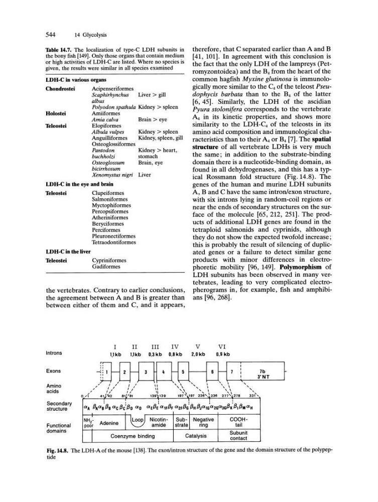
found in all dehydrogenases, and this has a typical Rossmann fold structure (Fig. 14.8). The
genes of the human and murine LDH subunits
A, Band C have the same intronlexon structure,
with six introns lying in random-coil regions or
near the ends of secondary structures on the surface of the molecule [65, 212, 251]. The products of additional LDH genes are found in the
tetraploid salmonids and cyprinids, although
they do not show the expected twofold increase;
this is probably the result of silencing of duplicated genes or a failure to detect similar gene
products with minor differences in electrophoretic mobility [96, 149]. Polymorpbism of
LDH subunits has been observed in many vertebrates, leading to very complicated electropherograms in, for example, fish and amphibians [96, 268].
V
2,Okb
" ,
6
VI
0,9 kb
,
I
7b
3'NT
, ,
,
0'1
41J'42
81~'81
139~139
, \
, \
\,
' \
,
I
,
I
197 '",,197 236' 236
" \
, I
, \
277'~278
, , ,
331 ,
Secondary
structure
Functional
domains
,
I
I
I
I
aA 13Aa a' 13a ae 13e: 13D aD ae13e' a1F13F a2F13G' 13H 13Ja 1G'a2Ga 3G13K' 131 13M a H
NH 2 • Adenine I ~ Nic~tin- SUb-II !Negative
COOHpoor
amide
strate
ring
tail
I
Coenzyme binding
Catalysis
Subunit
contact
Fig.14.8. The LDH-A of the mouse [138]. The exonlintron structure of the gene and the domain stru