

486
13 The Structure and Metabolism of Carbohydrates
egg shells of the platyhelminths, nematodes and
acanthocephalans; the tubes of the pogonophorans; the perisarc of the hydrozoans; the internal
skeleton of many hexacorallia; and the gemmules
of many sponges [72, 123,192]. Chitin appears to
be completely absent from the group of phyla
known as the Deuterostomia, i.e. echinoderms,
hemichordates and chordates; there is, however,
an unambiguous report of chitin in the gill arches
of the lanzelet Branchiostoma floridae. Chitin has
also been discovered in the arrow worms (chaetognathans), whose membership in the Deuterostomia is very doubtful [8].
Chitin is usually described as a linear ~(1-4)
polymer of N-acetylglucosamine but in many
cases, even those involving less destructive isolation methods, some sugars turn out to be nonacetylated. This probably involves the activity of
a chitin deacetylase, which is known in bacteria
but has never been directly demonstrated in animals. The chitin of insect cuticulae usually contains about 1-20 % de ace tyla ted residues. The
irregular abdominal expansion of the queen in
the termite species Macroterms estherae involves
deacetylation: the degree of de acetylation of the
abdominal cuticula increases from zero in the
winged sexually mature animal to over 90 % during egg production [158]. The chitin chain has a
conformation (ribbon conformation) similar to
that of the cellulose chain and, thus, can form fiba)
ot
b)
Chitin microfibril
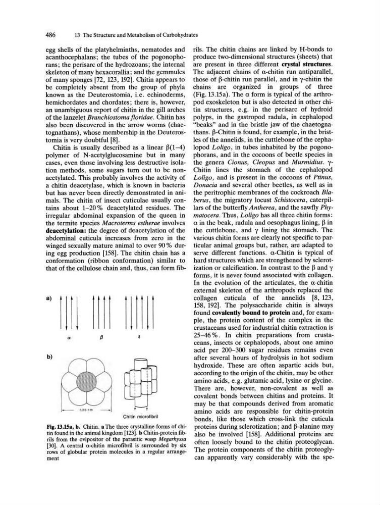
Fig.13.lSa, b. Chitin. a The three crystalline forms of chitin found in the animal kingdom [123]. b Chitin-protein fibrils from the ovipositor of the parasitic wasp Megarhyssa
[30]. A central a-chitin microfibril is surrounded by six
rows of globular protein molecules in a regular arrangement
rils. The chitin chains are linked by H-bonds to
produce two-dimensional structures (sheets) that
are present in three different crystal structures.
The adjacent chains of a-chitin run antiparallel,
those of ~-chitin run parallel, and in y-chitin the
chains are organized in groups of three
(Fig. 13.15a). The a form is typical of the arthropod exoskeleton but is also detected in other chitin structures, e.g. in the perisarc of hydroid
polyps, in the gastropod radula, in cephalopod
"beaks" and in the bristle jaw of the chaetognathans. ~-Chitin is found, for example, in the bristles of the annelids, in the cuttlebone of the cephalopod Loligo, in tubes inhabited by the pogo nophorans, and in the cocoons of beetle species in
the genera Cionus, Cleopus and Murmidius. yChitin lines the stomach of the cephalopod
Loligo, and is present in the cocoons of Ptinus,
Donacia and several other beetles, as well as in
the peri trophic membranes of the cockroach Blaberus, the migratory locust Schistocera, caterpillars of the butterfly Antherea, and the sawfly Phymatocera. Thus, Loligo has all three chitin forms:
a in the beak, radula and oesophagus lining, ~ in
the cuttlebone, and y lining the stomach. The
various chitin forms are clearly not specific to particular animal groups but, rather, are adapted to
serve different functions. a-Chitin is typical of
hard structures which are strengthened by sclerotization or calcification. In contrast to the ~ and y
forms, it is never found associated with collagen.
In the evolution of the articulates, the a-chitin
external skeleton of the arthropods replaced the
collagen cuticula of the annelids [8, 123,
158, 192]. The polysaccharide chitin is always
found covalently bound to protein and, for example, the protein content of the complex in the
crustaceans used for industrial chitin extraction is
25-46 % . In chitin preparations from crustaceans, insects or cephalopods, about one amino
acid per 200-300 sugar residues remains even
after several hours of hydrolysis in hot sodium
hydroxide. These are often aspartic acids but,
according to the origin of the chitin, may be other
amino acids, e.g. glutamic acid, lysine or glycine.
There are, however, non-covalent as well as
covalent bonds between chitins and proteins. It
may be that compounds derived from aromatic
amino acids are r~sponsible for chitin-protein
bonds, like those which cross-link the cuticula
proteins during sclerotization; and ~-alanine may
also be involved [158]. Additional proteins are
often loosely bound to the chitin proteoglycan.
The protein components of the chitin proteoglycan apparently vary considerably with the spe-