

sion of 3-hydroxykynurenine to 3-hydroxyanthranilic acid in the main pathway of tryptophan catabolism, and in Bombyx this enzyme
brings about the formation of anthranilic acid and
its conjugates from kynurenine [137]. Kynurenic
acid and xanthurenic acid are produced by amino
transfer from kynurenine or 3-hydroxykynurenine to 2-oxoglutarate, in proportions that
depend upon the species. The relevant enzymes
of Schistocerca gregaria and Musca domestica
have been examined in some detail.
12.5.5 Iodamino Acids
The synthesis of the thyroid hormone involves the
iodination of the tyrosine residues of a specific
protein, thyroglobulin, through the activity of a
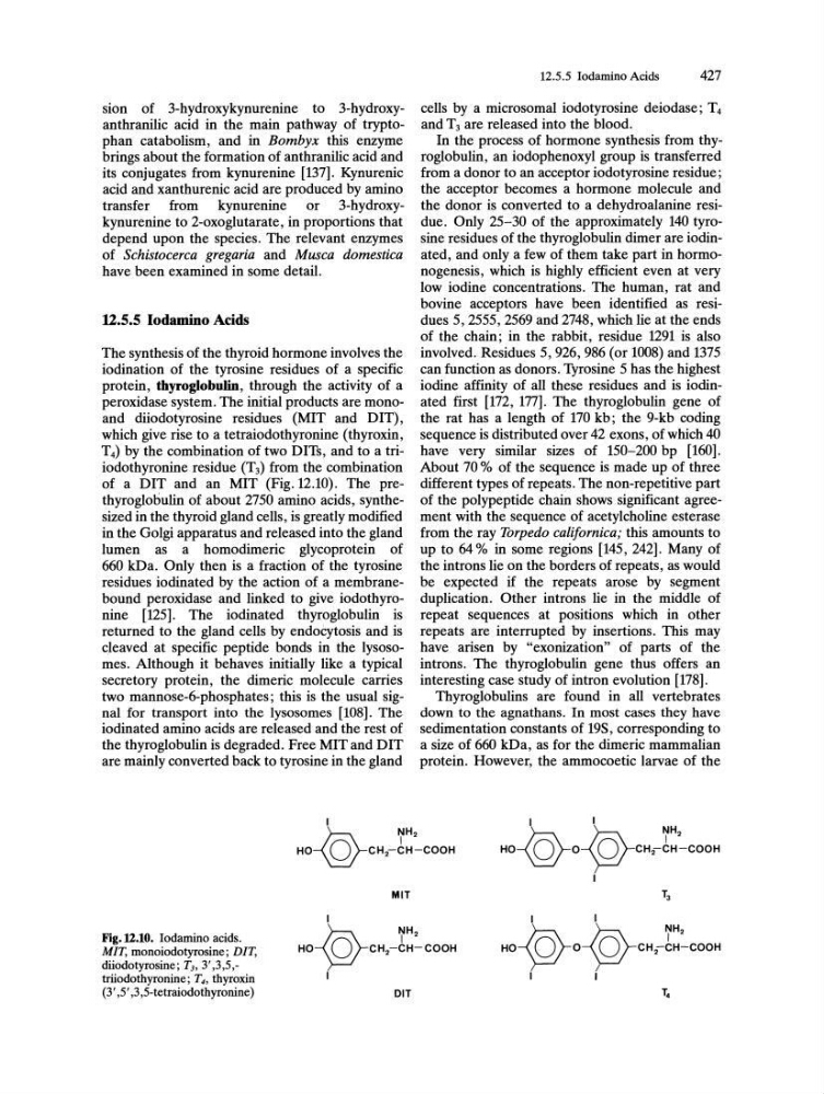
peroxidase system. The initial products are monoand diiodotyrosine residues (MIT and DIT) ,
which give rise to a tetraiodothyronine (thyroxin,
T4) by the combination of two DITs, and to a triiodothyronine residue (T3) from the combination
of a DIT and an MIT (Fig. 12.10). The prethyroglobulin of about 2750 amino acids, synthesized in the thyroid gland cells, is greatly modified
in the Golgi apparatus and released into the gland
lumen as a homodimeric glycoprotein of
660 kDa. Only then is a fraction of the tyrosine
residues iodinated by the action of a membranebound peroxidase and linked to give iodothyronine [125]. The iodinated thyroglobulin is
returned to the gland cells by endocytosis and is
cleaved at specific peptide bonds in the lysosomeso Although it behaves initially like a typical
secretory protein, the dimeric molecule carries
two mannose-6-phosphates; this is the usual signal for transport into the lysosomes [108]. The
iodinated amino acids are released and the rest of
the thyroglobulin is degraded. Free MIT and DIT
are mainly converted back to tyrosine in the gland
Fig.12.10. Iodamino acids.
MIT, monoiodotyrosine; DIT,
diiodotyrosine; T3 , 3' ,3,5,triiodothyronine; T4, thyroxin
(3',5' ,3,5-tetraiodothyronine)
MIT
OIT
12.5.5 Iodamino Acids
427
cells by a microsomal iodotyrosine deiodase; T 4
and T3 are released into the blood.
In the process of hormone synthesis from thyroglobulin, an iodophenoxyl group is transferred
from a donor to an acceptor iodotyrosine residue;
the acceptor becomes a hormone molecule and
the donor is converted to a dehydroalanine residue. Only 25-30 of the approximately 140 tyrosine residues of the thyroglobulin dimer are iodinated, and only a few of them take part in hormonogenesis, which is highly efficient even at very
low iodine concentrations. The human, rat and
bovine acceptors have been identified as residues 5,2555,2569 and 2748, which lie at the ends
of the chain; in the rabbit, residue 1291 is also
involved. Residues 5,926,986 (or 1008) and 1375
can function as donors. Tyrosine 5 has the highest
iodine affinity of all these residues and is iodinated first [172, 177]. The thyroglobulin gene of
the rat has a length of 170 kb; the 9-kb coding
sequence is distributed over 42 exons, of which 40
have very similar sizes of 150-200 bp [160].
About 70 % of the sequence is made up of three
different types of repeats. The non-repetitive part
of the polypeptide chain shows significant agreement with the sequence of acetylcholine esterase
from the ray Torpedo californica; this amounts to
up to 64 % in some regions [145, 242]. Many of
the introns lie on the borders of repeats, as would
be expected if the repeats arose by segment
duplication. Other introns lie in the middle of
repeat sequences at positions which in other
repeats are interrupted by insertions. This may
have arisen by "exonization" of parts of the
introns. The thyroglobulin gene thus offers an
interesting case study of intron evolution [178].
Thyroglobulins are found in all vertebrates
down to the agnathans. In most cases they have
sedimentation constants of 19S, corresponding to
a size of 660 kDa, as for the dimeric mammalian
protein. However, the ammocoetic larvae of the
brings about the formation of anthranilic acid and
its conjugates from kynurenine [137]. Kynurenic
acid and xanthurenic acid are produced by amino
transfer from kynurenine or 3-hydroxykynurenine to 2-oxoglutarate, in proportions that
depend upon the species. The relevant enzymes
of Schistocerca gregaria and Musca domestica
have been examined in some detail.
12.5.5 Iodamino Acids
The synthesis of the thyroid hormone involves the
iodination of the tyrosine residues of a specific
protein, thyroglobulin, through the activity of a
peroxidase system. The initial products are monoand diiodotyrosine residues (MIT and DIT) ,
which give rise to a tetraiodothyronine (thyroxin,
T4) by the combination of two DITs, and to a triiodothyronine residue (T3) from the combination
of a DIT and an MIT (Fig. 12.10). The prethyroglobulin of about 2750 amino acids, synthesized in the thyroid gland cells, is greatly modified
in the Golgi apparatus and released into the gland
lumen as a homodimeric glycoprotein of
660 kDa. Only then is a fraction of the tyrosine
residues iodinated by the action of a membranebound peroxidase and linked to give iodothyronine [125]. The iodinated thyroglobulin is
returned to the gland cells by endocytosis and is
cleaved at specific peptide bonds in the lysosomeso Although it behaves initially like a typical
secretory protein, the dimeric molecule carries
two mannose-6-phosphates; this is the usual signal for transport into the lysosomes [108]. The
iodinated amino acids are released and the rest of
the thyroglobulin is degraded. Free MIT and DIT
are mainly converted back to tyrosine in the gland
Fig.12.10. Iodamino acids.
MIT, monoiodotyrosine; DIT,
diiodotyrosine; T3 , 3' ,3,5,triiodothyronine; T4, thyroxin
(3',5' ,3,5-tetraiodothyronine)
MIT
OIT
12.5.5 Iodamino Acids
427
cells by a microsomal iodotyrosine deiodase; T 4
and T3 are released into the blood.
In the process of hormone synthesis from thyroglobulin, an iodophenoxyl group is transferred
from a donor to an acceptor iodotyrosine residue;
the acceptor becomes a hormone molecule and
the donor is converted to a dehydroalanine residue. Only 25-30 of the approximately 140 tyrosine residues of the thyroglobulin dimer are iodinated, and only a few of them take part in hormonogenesis, which is highly efficient even at very
low iodine concentrations. The human, rat and
bovine acceptors have been identified as residues 5,2555,2569 and 2748, which lie at the ends
of the chain; in the rabbit, residue 1291 is also
involved. Residues 5,926,986 (or 1008) and 1375
can function as donors. Tyrosine 5 has the highest
iodine affinity of all these residues and is iodinated first [172, 177]. The thyroglobulin gene of
the rat has a length of 170 kb; the 9-kb coding
sequence is distributed over 42 exons, of which 40
have very similar sizes of 150-200 bp [160].
About 70 % of the sequence is made up of three
different types of repeats. The non-repetitive part
of the polypeptide chain shows significant agreement with the sequence of acetylcholine esterase
from the ray Torpedo californica; this amounts to
up to 64 % in some regions [145, 242]. Many of
the introns lie on the borders of repeats, as would
be expected if the repeats arose by segment
duplication. Other introns lie in the middle of
repeat sequences at positions which in other
repeats are interrupted by insertions. This may
have arisen by "exonization" of parts of the
introns. The thyroglobulin gene thus offers an
interesting case study of intron evolution [178].
Thyroglobulins are found in all vertebrates
down to the agnathans. In most cases they have
sedimentation constants of 19S, corresponding to
a size of 660 kDa, as for the dimeric mammalian
protein. However, the ammocoetic larvae of the