

well as the sites responsible for the binding of
nidogenlentactin and for calcium-dependent
aggregation, lie on the short arms of the
B chains. The large globule at the C-terminus of
the A chains binds glycosaminoglycans [8]. The
sequences of the human and murine A chains,
with 3058-3060 amino acids, agree by 76 % and,
with the exception of the globular region, are significantly homologous to the B chains. The various regions of the human Bl (1765 amino acids)
and B2 (1576 amino acids) chains agree in their
sequences by only 16-40 %. The Bl gene includes 34 exons and the B2 gene only 28; of the
introns, only three occupy the same positions in
both genes [60,90]. Laminins have been
detected in various invertebrates, but only the
B chain of Drosophila melanogaster has been
sequenced. The B2 chain of the fly agrees at
40-41 % of positions with human and murine B2,
but at only 29-30 % with its own Bl chain or the
Bl of the mammals [18]. The basement membranes' of the trophoblasts from human placenta, of
striated skeletal muscle and of the Schwann cells
from the peripheral nervous system contain a
specific laminin made up of the polypeptide
merosin and the laminin chains Bl and B2. The
C-terminal region of merosin, as yet the only
characterized region of the molecule, shows 40 %
sequence similarity to the laminin A chain [30].
The laminin of the basement membranes is often
complexed with nidogen (entactin). Murine
nidogen is a dumb-bell-shaped molecule carrying
binding sites for cells, laminin and collagen IV.
The globular N- and C-terminal domains of nidogen consist of 641 and 328 amino acids, respectively, and the connecting rod domain has 248
amino acids [77]. Glutactin from the basement
membranes of Drosophila may be functionally
similar to nidogen but structurally it is very different; the N-terminal half of its sequence of
1023 amino acids resembles serine enzymes, but
11.1 Fibronectins
377
catalytic activity is absent; the C-terminal half
contains no fewer than 44 % glutamate and glutamine residues [93].
11.1 Fibronectins
Fibronectins are found in vertebrates as insoluble
structural proteins on the surface of fibroblasts
and other cells, in the connective tissue matrix,
and between the basement membrane and the
associated cells; they are also found in soluble
form as cell-adhesion factors in the blood plasma.
They are present at a much greater concentration
in embryonal tissues than in the adult, and are
thought to have an organizing function in tissue
formation. Only the structures of the human,
bovine and rat soluble forms are known in any
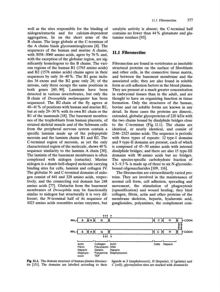
detail. In these cases the proteins are dimeric,
extended, globular glycoproteins of 220 kDa with
the two chains bound by disulphide bridges close
to the C-terminus (Fig. 11.1). The chains are
identical, or nearly identical, and consist of
2146-2325 amino acids. The sequence is periodic
with three types of repeats: 12 type-I domains
and 9 type-II domains are present, each of which
is composed of 45-50 amino acids with internal
disulphide bridges; and there are also 15 type-III
domains with 90 amino acids but no bridges.
The species-specific carbohydrate fraction of
4.5-9.5 % is made up of three to six N-glycosidicbound oligo saccharides [109, 116].
The fibronectins are extraordinarily varied proteins. They are involved in the maintenance of
normal cell form, cell adhesion, spreading and
movement, the stimulation of phagocytosis
(opsonification) and wound healing; they bind
collagen, fibrin, actin and other proteins of the
membrane skeleton, heparin, hyaluronic acid,
gangliosides, polyamines, the complement com' - -_ _ --I' L....' _ _ ~I '-, _ _ _ _ _ _ _ _ _ ---J' L...--....I ,-I _ _ ~
Actin
Fibrin
Heparin
Staphylococci
Collagen
Actin
Fibronectin DNA
Gelatin
Heparin
Polyamine Staphylococci
TEF
Cells Heparin
Fig.Il.I. The domain structure of human plasma fibronectin [131]. The domains are labelled according to their
ligands as S (staphylococci), H (heparin), G (gelatin) and
C (cell); glycosylation sites are marked with drumsticks
nidogenlentactin and for calcium-dependent
aggregation, lie on the short arms of the
B chains. The large globule at the C-terminus of
the A chains binds glycosaminoglycans [8]. The
sequences of the human and murine A chains,
with 3058-3060 amino acids, agree by 76 % and,
with the exception of the globular region, are significantly homologous to the B chains. The various regions of the human Bl (1765 amino acids)
and B2 (1576 amino acids) chains agree in their
sequences by only 16-40 %. The Bl gene includes 34 exons and the B2 gene only 28; of the
introns, only three occupy the same positions in
both genes [60,90]. Laminins have been
detected in various invertebrates, but only the
B chain of Drosophila melanogaster has been
sequenced. The B2 chain of the fly agrees at
40-41 % of positions with human and murine B2,
but at only 29-30 % with its own Bl chain or the
Bl of the mammals [18]. The basement membranes' of the trophoblasts from human placenta, of
striated skeletal muscle and of the Schwann cells
from the peripheral nervous system contain a
specific laminin made up of the polypeptide
merosin and the laminin chains Bl and B2. The
C-terminal region of merosin, as yet the only
characterized region of the molecule, shows 40 %
sequence similarity to the laminin A chain [30].
The laminin of the basement membranes is often
complexed with nidogen (entactin). Murine
nidogen is a dumb-bell-shaped molecule carrying
binding sites for cells, laminin and collagen IV.
The globular N- and C-terminal domains of nidogen consist of 641 and 328 amino acids, respectively, and the connecting rod domain has 248
amino acids [77]. Glutactin from the basement
membranes of Drosophila may be functionally
similar to nidogen but structurally it is very different; the N-terminal half of its sequence of
1023 amino acids resembles serine enzymes, but
11.1 Fibronectins
377
catalytic activity is absent; the C-terminal half
contains no fewer than 44 % glutamate and glutamine residues [93].
11.1 Fibronectins
Fibronectins are found in vertebrates as insoluble
structural proteins on the surface of fibroblasts
and other cells, in the connective tissue matrix,
and between the basement membrane and the
associated cells; they are also found in soluble
form as cell-adhesion factors in the blood plasma.
They are present at a much greater concentration
in embryonal tissues than in the adult, and are
thought to have an organizing function in tissue
formation. Only the structures of the human,
bovine and rat soluble forms are known in any
detail. In these cases the proteins are dimeric,
extended, globular glycoproteins of 220 kDa with
the two chains bound by disulphide bridges close
to the C-terminus (Fig. 11.1). The chains are
identical, or nearly identical, and consist of
2146-2325 amino acids. The sequence is periodic
with three types of repeats: 12 type-I domains
and 9 type-II domains are present, each of which
is composed of 45-50 amino acids with internal
disulphide bridges; and there are also 15 type-III
domains with 90 amino acids but no bridges.
The species-specific carbohydrate fraction of
4.5-9.5 % is made up of three to six N-glycosidicbound oligo saccharides [109, 116].
The fibronectins are extraordinarily varied proteins. They are involved in the maintenance of
normal cell form, cell adhesion, spreading and
movement, the stimulation of phagocytosis
(opsonification) and wound healing; they bind
collagen, fibrin, actin and other proteins of the
membrane skeleton, heparin, hyaluronic acid,
gangliosides, polyamines, the complement com' - -_ _ --I' L....' _ _ ~I '-, _ _ _ _ _ _ _ _ _ ---J' L...--....I ,-I _ _ ~
Actin
Fibrin
Heparin
Staphylococci
Collagen
Actin
Fibronectin DNA
Gelatin
Heparin
Polyamine Staphylococci
TEF
Cells Heparin
Fig.Il.I. The domain structure of human plasma fibronectin [131]. The domains are labelled according to their
ligands as S (staphylococci), H (heparin), G (gelatin) and
C (cell); glycosylation sites are marked with drumsticks