

358
10 Proteins of Muscle and the Cytoskeleton
cDNA; this protein is highly homologous to
mammalian keratins of type II [73].
The hard keratins of the mammals are complex
structures which contain other, unrelated proteins
in addition to cytokeratins. The best-investigated
example is sheep wool. The keratinocytes in the
hair follicles of the sheep produce filaments or fibrils from low-sulphur (LS) proteins that have a
mass of 45-58 kDa and only 5 % cysteine. In contrast to the skin cells, the filaments of the hairfollicle cells are embedded in a matrix of proteins
from two further families: the high-sulphur (HS)
proteins (11-26 kDa and up to 30% cysteine),
and the glycine/tyrosine-rich (HGT) proteins of
6-9 kDa and with up to 30 % glycine and 15 %
tyrosine. The compressibility of the keratin structures depends upon the proportion of matrix proteins. For example, the ratios of LS:HS:HGT
proteins in sheep are 62:29:9 in the wool,
76: 11 : 13 in horn and 79: 11 : 10 in the hooves [236].
The composition of mammalian hair, and hence its
mechanical properties, differs between species
and races, and is also dependent upon external
and internal conditions. The LS proteins are similar in amino acid sequence and tertiary structure
to the cytokeratins. In sheep wool, there are two
groups, each of four LS proteins, which apparently correspond to the types I and II of the cytokeratins [254]. Two HGTproteins and 18 HS proteins have been sequenced so far; they are not
related to each other or to the cytokeratins. The
genes of both protein families contain no introns.
The differences between the hard keratins of
the mammals and those of both reptiles and birds
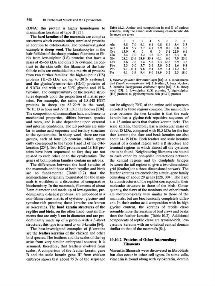
are so fundamental (Table 10.2) that the
nomenclature originally formulated for the mammals is worthless in a discussion of comparative
biochemistry. In the mammals, filaments of about
7-nm diameter and made up of low-cysteine, predominantly a-helical proteins, are embedded in a
non-filamentous matrix of cysteine-, glycine- and
tyrosine-rich proteins; these keratins are known
as a-keratins. The hard keratin structures of the
reptiles and birds, on the other hand, contain filaments that are only 3 nm in diameter and are predominantly made up of a protein with a B-sheet
structure; this type is termed cp- or B-keratin [84].
The best-investigated examples of B-keratins
are the feather keratins of the chicken and other
bird species. The feathers and the scales of the leg
arise from very similar embryonal sources; it is
assumed, therefore, that feathers evolved from
scales. A comparison of the feather keratin gene
B and the scale keratin gene III from chicken
embryos shows that about 75 % of the sequence
Thble 10.2. Amino acid composition in mol % of various
keratins. Only the amino acids showing characteristic differences are given
1
2
3
4
5
6
7
8
Ala
4.8 7.0 6.1 6.1 0.8 8.4 3.4 3.3
Asp
4.8 5.9 3.7 4.1 2.9 0.8 0.8 1.6
Cys
13.0
0
0
0
0
5.1 22.0 6.6
Glu
3.4 7.0 3.2 3.8 0.7 0.5 2.3
0
Gly
28.2 15.6 33.9 30.8 40.1 6.0 7.0 23.0
Leu
3.6 7.8 5.4 5.8 5.3 12.8 2.9 3.3
Phe
2.3 3.0 3.7 3.6 5.0 3.2 1.6 9.8
Pro
9.1 11.1 9.6 9.4 3.9 1.4 11.2 6.6
1Yr
4.1 3.9 9.4 9.0 18.9 3.2 2.3 18.0
1, Varanus gouldii; claw outer layer [84]; 2-4, Kookaburra
bird Dacelo novaeguineae [84]: 2, feather; 3, beak; 4, claw;
5, echidna Tachyglossus aculeatus; spine [84]; 6-8, sheep
wool [53]: 6, low-sulphur (LS) protein; 7, high-sulphur
(HS) protein; 8, glycine/tyrosine-rich (HGT) protein
can be aligned; 70 % of the amino acid sequences
encoded by these regions coincide. The main difference between the two keratins is that the scale
keratin has a glycine-rich repetitive sequence of
4 X 13 amino acids that feather keratin lacks. The
scale keratin, therefore, has a molecular mass of
about 15 kDa, compared with 10.5 kDa for the feather keratin; the claw and beak keratins are also
about 14-15 kDa. Both feather and scale keratins
consist of a central region with a B structure and
terminal regions in which almost all the cysteines
are to be found. Neighbouring filaments are bound
to each other by non-polar interactions between
the central regions and by disulphide bridges
between the tail regions to give a two-dimensional
grid (feather) or a three-dimensional network. The
feather keratins are encoded by a multi-gene family
consisting of about 20 genes [228, 304]. The hard
keratin structures of the reptiles correspond in their
molecular structure to those of the birds. Consequently, the claws of the monitors and other lizards
are morphologically very similar to those of the
mammals, but are biochemically completely different. In their amino acid composition with its high
glycine content, the keratins of reptile claws
resemble more the keratins of bird claws and beaks
than the feather keratins (Table 10.2). Additional
components of reptile claws are tyrosine-rich, lowcysteine keratins with an a-helical central domain
similar to that of the mammals [84].
10.10.2 Proteins of Other Intermediary
Filaments
Vimentin filaments were discovered in fibroblasts
but also occur in other cell types. In some cells,
vimentin is found along with cytokeratin, desmin
10 Proteins of Muscle and the Cytoskeleton
cDNA; this protein is highly homologous to
mammalian keratins of type II [73].
The hard keratins of the mammals are complex
structures which contain other, unrelated proteins
in addition to cytokeratins. The best-investigated
example is sheep wool. The keratinocytes in the
hair follicles of the sheep produce filaments or fibrils from low-sulphur (LS) proteins that have a
mass of 45-58 kDa and only 5 % cysteine. In contrast to the skin cells, the filaments of the hairfollicle cells are embedded in a matrix of proteins
from two further families: the high-sulphur (HS)
proteins (11-26 kDa and up to 30% cysteine),
and the glycine/tyrosine-rich (HGT) proteins of
6-9 kDa and with up to 30 % glycine and 15 %
tyrosine. The compressibility of the keratin structures depends upon the proportion of matrix proteins. For example, the ratios of LS:HS:HGT
proteins in sheep are 62:29:9 in the wool,
76: 11 : 13 in horn and 79: 11 : 10 in the hooves [236].
The composition of mammalian hair, and hence its
mechanical properties, differs between species
and races, and is also dependent upon external
and internal conditions. The LS proteins are similar in amino acid sequence and tertiary structure
to the cytokeratins. In sheep wool, there are two
groups, each of four LS proteins, which apparently correspond to the types I and II of the cytokeratins [254]. Two HGTproteins and 18 HS proteins have been sequenced so far; they are not
related to each other or to the cytokeratins. The
genes of both protein families contain no introns.
The differences between the hard keratins of
the mammals and those of both reptiles and birds
are so fundamental (Table 10.2) that the
nomenclature originally formulated for the mammals is worthless in a discussion of comparative
biochemistry. In the mammals, filaments of about
7-nm diameter and made up of low-cysteine, predominantly a-helical proteins, are embedded in a
non-filamentous matrix of cysteine-, glycine- and
tyrosine-rich proteins; these keratins are known
as a-keratins. The hard keratin structures of the
reptiles and birds, on the other hand, contain filaments that are only 3 nm in diameter and are predominantly made up of a protein with a B-sheet
structure; this type is termed cp- or B-keratin [84].
The best-investigated examples of B-keratins
are the feather keratins of the chicken and other
bird species. The feathers and the scales of the leg
arise from very similar embryonal sources; it is
assumed, therefore, that feathers evolved from
scales. A comparison of the feather keratin gene
B and the scale keratin gene III from chicken
embryos shows that about 75 % of the sequence
Thble 10.2. Amino acid composition in mol % of various
keratins. Only the amino acids showing characteristic differences are given
1
2
3
4
5
6
7
8
Ala
4.8 7.0 6.1 6.1 0.8 8.4 3.4 3.3
Asp
4.8 5.9 3.7 4.1 2.9 0.8 0.8 1.6
Cys
13.0
0
0
0
0
5.1 22.0 6.6
Glu
3.4 7.0 3.2 3.8 0.7 0.5 2.3
0
Gly
28.2 15.6 33.9 30.8 40.1 6.0 7.0 23.0
Leu
3.6 7.8 5.4 5.8 5.3 12.8 2.9 3.3
Phe
2.3 3.0 3.7 3.6 5.0 3.2 1.6 9.8
Pro
9.1 11.1 9.6 9.4 3.9 1.4 11.2 6.6
1Yr
4.1 3.9 9.4 9.0 18.9 3.2 2.3 18.0
1, Varanus gouldii; claw outer layer [84]; 2-4, Kookaburra
bird Dacelo novaeguineae [84]: 2, feather; 3, beak; 4, claw;
5, echidna Tachyglossus aculeatus; spine [84]; 6-8, sheep
wool [53]: 6, low-sulphur (LS) protein; 7, high-sulphur
(HS) protein; 8, glycine/tyrosine-rich (HGT) protein
can be aligned; 70 % of the amino acid sequences
encoded by these regions coincide. The main difference between the two keratins is that the scale
keratin has a glycine-rich repetitive sequence of
4 X 13 amino acids that feather keratin lacks. The
scale keratin, therefore, has a molecular mass of
about 15 kDa, compared with 10.5 kDa for the feather keratin; the claw and beak keratins are also
about 14-15 kDa. Both feather and scale keratins
consist of a central region with a B structure and
terminal regions in which almost all the cysteines
are to be found. Neighbouring filaments are bound
to each other by non-polar interactions between
the central regions and by disulphide bridges
between the tail regions to give a two-dimensional
grid (feather) or a three-dimensional network. The
feather keratins are encoded by a multi-gene family
consisting of about 20 genes [228, 304]. The hard
keratin structures of the reptiles correspond in their
molecular structure to those of the birds. Consequently, the claws of the monitors and other lizards
are morphologically very similar to those of the
mammals, but are biochemically completely different. In their amino acid composition with its high
glycine content, the keratins of reptile claws
resemble more the keratins of bird claws and beaks
than the feather keratins (Table 10.2). Additional
components of reptile claws are tyrosine-rich, lowcysteine keratins with an a-helical central domain
similar to that of the mammals [84].
10.10.2 Proteins of Other Intermediary
Filaments
Vimentin filaments were discovered in fibroblasts
but also occur in other cell types. In some cells,
vimentin is found along with cytokeratin, desmin