

354
10 Proteins of Muscle and the Cytoskeleton
Tubule
Central tubules
larger and more complex than the head region of
the myosin molecule. Their large size has hindered analysis by the usual methods of protein
chemistry. The outer dynein arms of the axonemes and the inner dynein arms, which may be
present in two different forms, have basically the
same structure but a very different polypeptide
composition. The inner arms are not very well
described, the best-known being the outer dynein
arms from the cilia of Tetrahymena, the flagella of
Chlamydomonas and the tails of sperm from sea
urchins and trout [78, 83, 179]. Electron microscope pictures of the outer-arm dyneins show two
or three globular heads arising from a common
point on flexible stems. The number of heads corresponds to the number of heavy chains (two in
sperm tails, and three in Chlamydomonas and
Tetrahymena). In a beating axoneme, a gliding
movement between the outer doublet tubules is
translated by elastic elements into a transverse
bending movement. An ATP-driven bridge cycle
occurs between the dynein arm on the A tubule of
a doublet and the B tubule of the neighbouring
doublet. The heavy chains in Tetrahymena may
carry up to six phosphate residues, with the phosphorylation presumably having some regulatory
function [36].
Cytoplasmic dyneins appear to be ubiquitous,
even occurring in Caenorhabditis, which, like all
nematodes, has no cilia or flagella. The dyneins
detected in the cytoplasm of sea urchin eggs probably represent, on the one hand, the maternal
contribution to cilia formation in the larva, but
perhaps also have egg-specific functions such as
cytoplasmic movement. A further microtubule
Outer dynein arm
Inner dynein arm
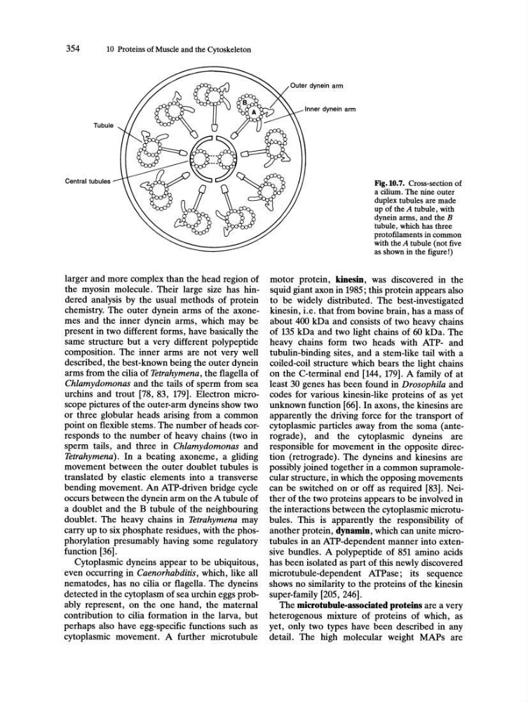
Fig.10.7. Cross-section of
a cilium. The nine outer
duplex tubules are made
up of the A tubule, with
dynein arms, and the B
tubule, which has three
proto filaments in common
with theA tubule (not five
as shown in the figure!)
motor protein, kinesin, was discovered in the
squid giant axon in 1985; this protein appears also
to be widely distributed. The best-investigated
kinesin, i.e. that from bovine brain, has a mass of
about 400 kDa and consists of two heavy chains
of 135 kDa and two light chains of 60 kDa. The
heavy chains form two heads with ATP- and
tubulin-binding sites, and a stem-like tail with a
coiled-coil structure which bears the light chains
on the C-terminal end [144, 179]. A family of at
least 30 genes has been found in Drosophila and
codes for various kinesin-like proteins of as yet
unknown function [66]. In axons, the kinesins are
apparently the driving force for the transport of
cytoplasmic particles away from the soma (anterograde), and the cytoplasmic dyneins are
responsible for movement in the opposite direction (retrograde). The dyneins and kinesins are
possibly joined together in a common supramolecular structure, in which the opposing movements
can be switched on or off as required [83]. Neither of the two proteins appears to be involved in
the interactions between the cytoplasmic microtubuIes. This is apparently the responsibility of
another protein, dynamin, which can unite microtubules in an ATP-dependent manner into extensive bundles. A polypeptide of 851 amino acids
has been isolated as part of this newly discovered
microtubule-dependent ATPase; its sequence
shows no similarity to the proteins of the kinesin
super-family [205, 246].
The microtubule-associated proteins are a very
heterogenous mixture of proteins of which, as
yet, only two types have been described in any
detail. The high molecular weight MAPs are
10 Proteins of Muscle and the Cytoskeleton
Tubule
Central tubules
larger and more complex than the head region of
the myosin molecule. Their large size has hindered analysis by the usual methods of protein
chemistry. The outer dynein arms of the axonemes and the inner dynein arms, which may be
present in two different forms, have basically the
same structure but a very different polypeptide
composition. The inner arms are not very well
described, the best-known being the outer dynein
arms from the cilia of Tetrahymena, the flagella of
Chlamydomonas and the tails of sperm from sea
urchins and trout [78, 83, 179]. Electron microscope pictures of the outer-arm dyneins show two
or three globular heads arising from a common
point on flexible stems. The number of heads corresponds to the number of heavy chains (two in
sperm tails, and three in Chlamydomonas and
Tetrahymena). In a beating axoneme, a gliding
movement between the outer doublet tubules is
translated by elastic elements into a transverse
bending movement. An ATP-driven bridge cycle
occurs between the dynein arm on the A tubule of
a doublet and the B tubule of the neighbouring
doublet. The heavy chains in Tetrahymena may
carry up to six phosphate residues, with the phosphorylation presumably having some regulatory
function [36].
Cytoplasmic dyneins appear to be ubiquitous,
even occurring in Caenorhabditis, which, like all
nematodes, has no cilia or flagella. The dyneins
detected in the cytoplasm of sea urchin eggs probably represent, on the one hand, the maternal
contribution to cilia formation in the larva, but
perhaps also have egg-specific functions such as
cytoplasmic movement. A further microtubule
Outer dynein arm
Inner dynein arm
Fig.10.7. Cross-section of
a cilium. The nine outer
duplex tubules are made
up of the A tubule, with
dynein arms, and the B
tubule, which has three
proto filaments in common
with theA tubule (not five
as shown in the figure!)
motor protein, kinesin, was discovered in the
squid giant axon in 1985; this protein appears also
to be widely distributed. The best-investigated
kinesin, i.e. that from bovine brain, has a mass of
about 400 kDa and consists of two heavy chains
of 135 kDa and two light chains of 60 kDa. The
heavy chains form two heads with ATP- and
tubulin-binding sites, and a stem-like tail with a
coiled-coil structure which bears the light chains
on the C-terminal end [144, 179]. A family of at
least 30 genes has been found in Drosophila and
codes for various kinesin-like proteins of as yet
unknown function [66]. In axons, the kinesins are
apparently the driving force for the transport of
cytoplasmic particles away from the soma (anterograde), and the cytoplasmic dyneins are
responsible for movement in the opposite direction (retrograde). The dyneins and kinesins are
possibly joined together in a common supramolecular structure, in which the opposing movements
can be switched on or off as required [83]. Neither of the two proteins appears to be involved in
the interactions between the cytoplasmic microtubuIes. This is apparently the responsibility of
another protein, dynamin, which can unite microtubules in an ATP-dependent manner into extensive bundles. A polypeptide of 851 amino acids
has been isolated as part of this newly discovered
microtubule-dependent ATPase; its sequence
shows no similarity to the proteins of the kinesin
super-family [205, 246].
The microtubule-associated proteins are a very
heterogenous mixture of proteins of which, as
yet, only two types have been described in any
detail. The high molecular weight MAPs are