

332
10 Proteins of Muscle and the Cytoskeleton
nal diameter 25 nm, internal diameter 15 nm) and
the intermediary filaments (with a diameter of
about 10 nm). The microtubules are made up
mostly of tubulin but can also bind other proteins
(the microtubule-associated proteins). The movement of flagella and cilia is the result of interaction between the microtubules and the ATPhydrolysing dynein. The composition of the intermediary filaments varies markedly with the cell
type, but the constituent proteins all belong to the
same super-family and present a particularly interesting example of adaptive evolution. They
include the cytokeratins of epithelial cells, the
vimentins from cells of mesenchyme origin, the
desmins of muscles, the neurofilament proteins,
and the acidic protein of the glial filaments
(GFAP). It has recently been demonstrated that
lamins A and C from the nuclear membrane are
homologous to proteins of the intermediary filaments. Specific differentiated cells contain
filament-like structures which do not conform to
any of the three general types (microfilaments,
intermediary filaments, microtubules), e.g. the
connectinltitin network of the myofibrils, the
spasmonemes in the stalk of the peritrichous ciliate Vorticella, or the myonemes of Stentor [244].
The spermatozoa of the nematode Caenorhabditis
elegans have no myosin and only a very little actin
and tubulin (0.02 % of the total protein), but contain large amounts (15 %) of specific basic proteins (main sperm proteins, MSPs) of 15 kDa; these
are mainly located in the pseudopodium and are
therefore probably involved in the movement of
these aberrant sex cells. Of the more than 30 MSP
genes present, at least three are expressed [28].
The components of the cytoskeleton are
involved in complex interactions with each other
and with the plasma membrane, and beyond this
with components of the extracellular matrix, such
as collagen, fibronectin and laminin. The
mechanical strength of the plasma membrane
relies upon a membrane skeleton of specific structural proteins; this is especially well developed in
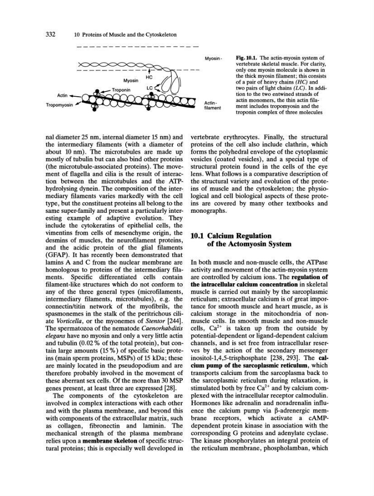
Actinfilament
Fig.IO.I. The actin-myosin system of
vertebrate skeletal muscle. For clarity,
only one myosin molecule is shown in
the thick myosin filament; this consists
of a pair of heavy chains (HC) and
two pairs of light chains (LC). In addition to the two entwined strands of
actin monomers, the thin actin filament includes tropomyosin and the
troponin complex of three molecules
vertebrate erythrocytes. Finally, the structural
proteins of the cell also include clathrin, which
forms the polyhedral envelope of the cytoplasmic
vesicles (coated vesicles), and a special type of
structural protein found in the cells of the eye
lens. What follows is a comparative description of
the structural variety and evolution of the proteins of muscle and the cytoskeleton; the physiological and cell biological aspects of these proteins are covered by many other textbooks and
monographs.
10.1 Calcium Regulation
of the Actomyosin System
In both muscle and non-muscle cells, the ATPase
activity and movement of the actin-myosin system
are controlled by calcium ions. The regulation of
the intracellular calcium concentration in skeletal
muscle is carried out mainly by the sarcoplasmic
reticulum; extracellular calcium is of great importance for smooth muscle and heart muscle, as is
calcium storage in the mitochondria of nonmuscle cells. In smooth muscle and non-muscle
cells, Ca 2 + is taken up from the outside by
potential-dependent or ligand-dependent calcium
channels, and is set free from intracellular reserves by the action of the secondary messenger
inositol-l,4,5-trisphosphate [238, 293]. The calcium pump of the sarcoplasmic reticulum, which
transports calcium from the sarcoplasma back to
the sarcoplasmic reticulum during relaxation, is
stimulated both by free Ca2+ and by calcium complexed with the intracellular receptor calmodulin.
Hormones like adrenalin and noradrenalin influence the calcium pump via ~-adrenergic membrane receptors, which activate a cAMPdependent protein kinase in association with the
corresponding G proteins and adenylate cyclase.
The kinase phosphorylates an integral protein of
the reticulum membrane, phospholamban, which
10 Proteins of Muscle and the Cytoskeleton
nal diameter 25 nm, internal diameter 15 nm) and
the intermediary filaments (with a diameter of
about 10 nm). The microtubules are made up
mostly of tubulin but can also bind other proteins
(the microtubule-associated proteins). The movement of flagella and cilia is the result of interaction between the microtubules and the ATPhydrolysing dynein. The composition of the intermediary filaments varies markedly with the cell
type, but the constituent proteins all belong to the
same super-family and present a particularly interesting example of adaptive evolution. They
include the cytokeratins of epithelial cells, the
vimentins from cells of mesenchyme origin, the
desmins of muscles, the neurofilament proteins,
and the acidic protein of the glial filaments
(GFAP). It has recently been demonstrated that
lamins A and C from the nuclear membrane are
homologous to proteins of the intermediary filaments. Specific differentiated cells contain
filament-like structures which do not conform to
any of the three general types (microfilaments,
intermediary filaments, microtubules), e.g. the
connectinltitin network of the myofibrils, the
spasmonemes in the stalk of the peritrichous ciliate Vorticella, or the myonemes of Stentor [244].
The spermatozoa of the nematode Caenorhabditis
elegans have no myosin and only a very little actin
and tubulin (0.02 % of the total protein), but contain large amounts (15 %) of specific basic proteins (main sperm proteins, MSPs) of 15 kDa; these
are mainly located in the pseudopodium and are
therefore probably involved in the movement of
these aberrant sex cells. Of the more than 30 MSP
genes present, at least three are expressed [28].
The components of the cytoskeleton are
involved in complex interactions with each other
and with the plasma membrane, and beyond this
with components of the extracellular matrix, such
as collagen, fibronectin and laminin. The
mechanical strength of the plasma membrane
relies upon a membrane skeleton of specific structural proteins; this is especially well developed in
Actinfilament
Fig.IO.I. The actin-myosin system of
vertebrate skeletal muscle. For clarity,
only one myosin molecule is shown in
the thick myosin filament; this consists
of a pair of heavy chains (HC) and
two pairs of light chains (LC). In addition to the two entwined strands of
actin monomers, the thin actin filament includes tropomyosin and the
troponin complex of three molecules
vertebrate erythrocytes. Finally, the structural
proteins of the cell also include clathrin, which
forms the polyhedral envelope of the cytoplasmic
vesicles (coated vesicles), and a special type of
structural protein found in the cells of the eye
lens. What follows is a comparative description of
the structural variety and evolution of the proteins of muscle and the cytoskeleton; the physiological and cell biological aspects of these proteins are covered by many other textbooks and
monographs.
10.1 Calcium Regulation
of the Actomyosin System
In both muscle and non-muscle cells, the ATPase
activity and movement of the actin-myosin system
are controlled by calcium ions. The regulation of
the intracellular calcium concentration in skeletal
muscle is carried out mainly by the sarcoplasmic
reticulum; extracellular calcium is of great importance for smooth muscle and heart muscle, as is
calcium storage in the mitochondria of nonmuscle cells. In smooth muscle and non-muscle
cells, Ca 2 + is taken up from the outside by
potential-dependent or ligand-dependent calcium
channels, and is set free from intracellular reserves by the action of the secondary messenger
inositol-l,4,5-trisphosphate [238, 293]. The calcium pump of the sarcoplasmic reticulum, which
transports calcium from the sarcoplasma back to
the sarcoplasmic reticulum during relaxation, is
stimulated both by free Ca2+ and by calcium complexed with the intracellular receptor calmodulin.
Hormones like adrenalin and noradrenalin influence the calcium pump via ~-adrenergic membrane receptors, which activate a cAMPdependent protein kinase in association with the
corresponding G proteins and adenylate cyclase.
The kinase phosphorylates an integral protein of
the reticulum membrane, phospholamban, which