

Chapter 3 Seagrass Morphology, Anatomy, and Ultrastructure
63
A
B
D
C
E
G
F
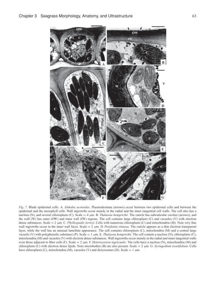
Fig. 7. Blade epidermal cells. A. Enhalus acoroides. Plasmodesmata (arrows) occur between two epidermal cells and between the
epidermal and the mesophyll cells. Wall ingrowths occur mainly in the radial and the inner tangential cell walls. The cell also has a
nucleus (N), and several chloroplasts (C). Scale = 4 µm. B. Thalassia hemprichii. The cuticle has subcuticular cavities (arrows), and
the wall (W) has outer (OW) and inner wall (IW) regions. The cell contains large chloroplasts (C) and vacuoles (V) with electron
dense substances. Scale = 2 µm. C. Phyllospadix torreyi. Cells with numerous chloroplasts (C) and mitochondria (M). Note very fine
wall ingrowths occur in the inner wall faces. Scale = 2 µm. D. Posidonia sinuosa. The cuticle appears as a thin electron transparent
layer, while the wall has an unusual lamellate appearance. The cell contains chloroplasts (C), mitochondria (M) and a central large
vacuole (V) with polyphenolic substance (P). Scale = 1 µm. E. Thalassia hemprichii. The cell contain a nucleus (N), chloroplasts (C),
mitochondria (M) and vacuoles (V) with electron dense substances. Wall ingrowths occur mainly in the radial and inner tangential walls
even those adjacent to fiber cells (F). Scale = 2 µm. F. Heterozostera nigricaulis. The cells have a nucleus (N), mitochondria (M) and
chloroplasts (C) with electron dense lipids. Note microbodies (B) are also present. Scale = 2 µm. G. Syringodium isoetifolium. Cells
have chloroplasts (C), mitochondria (M), vacuoles (V) and dictyosomes (D). Scale = 1 µm.
63
A
B
D
C
E
G
F
Fig. 7. Blade epidermal cells. A. Enhalus acoroides. Plasmodesmata (arrows) occur between two epidermal cells and between the
epidermal and the mesophyll cells. Wall ingrowths occur mainly in the radial and the inner tangential cell walls. The cell also has a
nucleus (N), and several chloroplasts (C). Scale = 4 µm. B. Thalassia hemprichii. The cuticle has subcuticular cavities (arrows), and
the wall (W) has outer (OW) and inner wall (IW) regions. The cell contains large chloroplasts (C) and vacuoles (V) with electron
dense substances. Scale = 2 µm. C. Phyllospadix torreyi. Cells with numerous chloroplasts (C) and mitochondria (M). Note very fine
wall ingrowths occur in the inner wall faces. Scale = 2 µm. D. Posidonia sinuosa. The cuticle appears as a thin electron transparent
layer, while the wall has an unusual lamellate appearance. The cell contains chloroplasts (C), mitochondria (M) and a central large
vacuole (V) with polyphenolic substance (P). Scale = 1 µm. E. Thalassia hemprichii. The cell contain a nucleus (N), chloroplasts (C),
mitochondria (M) and vacuoles (V) with electron dense substances. Wall ingrowths occur mainly in the radial and inner tangential walls
even those adjacent to fiber cells (F). Scale = 2 µm. F. Heterozostera nigricaulis. The cells have a nucleus (N), mitochondria (M) and
chloroplasts (C) with electron dense lipids. Note microbodies (B) are also present. Scale = 2 µm. G. Syringodium isoetifolium. Cells
have chloroplasts (C), mitochondria (M), vacuoles (V) and dictyosomes (D). Scale = 1 µm.