

Chapter 3 Seagrass Morphology, Anatomy, and Ultrastructure
61
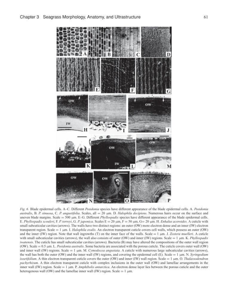
Fig. 6. Blade epidermal cells. A–C. Different Posidonia species have different appearance of the blade epidermal cells. A. Posidonia
australis, B. P. sinuosa, C. P. angustifolia. Scales, all = 20 µm. D. Halophila decipiens. Numerous hairs occur on the surface and
uneven blade margins. Scale = 500 µm. E–G. Different Phyllospadix species have different appearance of the blade epidermal cells.
E. Phyllospadix scouleri, F. P. torreyi, G. P. japonica. Scales E = 20 µm, F = 50 µm, G= 20 µm. H. Enhalus acoroides. A cuticle with
small subcuticular cavities (arrows). The walls have two distinct regions: an outer (OW) more electron dense and an inner (IW) electron
transparent region. Scale = 1 µm. I. Halophila ovalis. An electron transparent cuticle covers cell walls, which possess an outer (OW)
and the inner (IW) region. Note that wall ingrowths (T) on the inner face of the walls. Scale = 1 µm. J. Zostera muelleri. A cuticle
with small subcuticular cavities (arrows), the wall also consists of outer (OW) and inner (IW) regions. Scale = 1 µm. K. Phyllospadix
iwatensis. The cuticle has small subcuticular cavities (arrows). Bacteria (B) may have altered the compositions of the outer wall region
(OW). Scale = 0.5 µm. L. Posidonia australis. Some bacteria are associated with the porous cuticle. The cuticle covers outer wall (OW)
and inner wall (IW) regions. Scale = 1 µm. M. Cymodocea angustata. A cuticle with numerous large subcuticular cavities (arrows),
the wall has both the outer (OW) and the inner wall (IW) regions, and covering the epidermal cell (E). Scale = 1 µm. N. Syringodium
isoetifolium. A thin electron transparent cuticle covers the outer (OW) and inner (IW) wall region. Scale = 1 µm. O. Thalassodendron
pachyrhizum. A thin electron transparent cuticle with complex inclusions in the outer wall (OW) and lamellae arrangements in the
inner wall (IW) region. Scale = 1 µm. P. Amphibolis antarctica. An electron dense layer lies between the porous cuticle and the outer
heterogenous wall (OW) and the lamellae inner wall (IW) region. Scale = 1 µm.
61
Fig. 6. Blade epidermal cells. A–C. Different Posidonia species have different appearance of the blade epidermal cells. A. Posidonia
australis, B. P. sinuosa, C. P. angustifolia. Scales, all = 20 µm. D. Halophila decipiens. Numerous hairs occur on the surface and
uneven blade margins. Scale = 500 µm. E–G. Different Phyllospadix species have different appearance of the blade epidermal cells.
E. Phyllospadix scouleri, F. P. torreyi, G. P. japonica. Scales E = 20 µm, F = 50 µm, G= 20 µm. H. Enhalus acoroides. A cuticle with
small subcuticular cavities (arrows). The walls have two distinct regions: an outer (OW) more electron dense and an inner (IW) electron
transparent region. Scale = 1 µm. I. Halophila ovalis. An electron transparent cuticle covers cell walls, which possess an outer (OW)
and the inner (IW) region. Note that wall ingrowths (T) on the inner face of the walls. Scale = 1 µm. J. Zostera muelleri. A cuticle
with small subcuticular cavities (arrows), the wall also consists of outer (OW) and inner (IW) regions. Scale = 1 µm. K. Phyllospadix
iwatensis. The cuticle has small subcuticular cavities (arrows). Bacteria (B) may have altered the compositions of the outer wall region
(OW). Scale = 0.5 µm. L. Posidonia australis. Some bacteria are associated with the porous cuticle. The cuticle covers outer wall (OW)
and inner wall (IW) regions. Scale = 1 µm. M. Cymodocea angustata. A cuticle with numerous large subcuticular cavities (arrows),
the wall has both the outer (OW) and the inner wall (IW) regions, and covering the epidermal cell (E). Scale = 1 µm. N. Syringodium
isoetifolium. A thin electron transparent cuticle covers the outer (OW) and inner (IW) wall region. Scale = 1 µm. O. Thalassodendron
pachyrhizum. A thin electron transparent cuticle with complex inclusions in the outer wall (OW) and lamellae arrangements in the
inner wall (IW) region. Scale = 1 µm. P. Amphibolis antarctica. An electron dense layer lies between the porous cuticle and the outer
heterogenous wall (OW) and the lamellae inner wall (IW) region. Scale = 1 µm.