

Chapter 3 Seagrass Morphology, Anatomy, and Ultrastructure
55
A
B
C
D
E
F
G
H
J
I
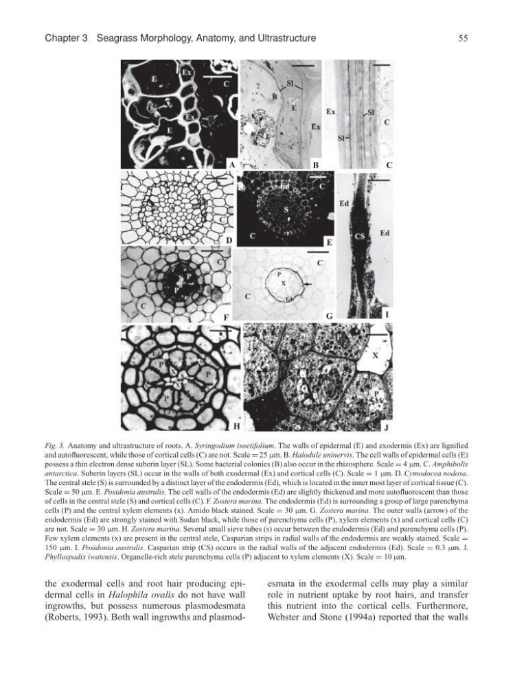
Fig. 3. Anatomy and ultrastructure of roots. A. Syringodium isoetifolium. The walls of epidermal (E) and exodermis (Ex) are lignified
and autofluorescent, while those of cortical cells (C) are not. Scale = 25 µm. B. Halodule uninervis. The cell walls of epidermal cells (E)
possess a thin electron dense suberin layer (SL). Some bacterial colonies (B) also occur in the rhizosphere. Scale = 4 µm. C. Amphibolis
antarctica. Suberin layers (SL) occur in the walls of both exodermal (Ex) and cortical cells (C). Scale = 1 µm. D. Cymodocea nodosa.
The central stele (S) is surrounded by a distinct layer of the endodermis (Ed), which is located in the inner most layer of cortical tissue (C).
Scale = 50 µm. E. Posidonia australis. The cell walls of the endodermis (Ed) are slightly thickened and more autofluorescent than those
of cells in the central stele (S) and cortical cells (C). F. Zostera marina. The endodermis (Ed) is surrounding a group of large parenchyma
cells (P) and the central xylem elements (x). Amido black stained. Scale = 30 µm. G. Zostera marina. The outer walls (arrow) of the
endodermis (Ed) are strongly stained with Sudan black, while those of parenchyma cells (P), xylem elements (x) and cortical cells (C)
are not. Scale = 30 µm. H. Zostera marina. Several small sieve tubes (s) occur between the endodermis (Ed) and parenchyma cells (P).
Few xylem elements (x) are present in the central stele, Casparian strips in radial walls of the endodermis are weakly stained. Scale =
150 µm. I. Posidonia australis. Casparian strip (CS) occurs in the radial walls of the adjacent endodermis (Ed). Scale = 0.3 µm. J.
Phyllospadix iwatensis. Organelle-rich stele parenchyma cells (P) adjacent to xylem elements (X). Scale = 10 µm.
the exodermal cells and root hair producing epidermal cells in Halophila ovalis do not have wall
ingrowths, but possess numerous plasmodesmata
(Roberts, 1993). Both wall ingrowths and plasmodesmata in the exodermal cells may play a similar
role in nutrient uptake by root hairs, and transfer
this nutrient into the cortical cells. Furthermore,
Webster and Stone (1994a) reported that the walls
55
A
B
C
D
E
F
G
H
J
I
Fig. 3. Anatomy and ultrastructure of roots. A. Syringodium isoetifolium. The walls of epidermal (E) and exodermis (Ex) are lignified
and autofluorescent, while those of cortical cells (C) are not. Scale = 25 µm. B. Halodule uninervis. The cell walls of epidermal cells (E)
possess a thin electron dense suberin layer (SL). Some bacterial colonies (B) also occur in the rhizosphere. Scale = 4 µm. C. Amphibolis
antarctica. Suberin layers (SL) occur in the walls of both exodermal (Ex) and cortical cells (C). Scale = 1 µm. D. Cymodocea nodosa.
The central stele (S) is surrounded by a distinct layer of the endodermis (Ed), which is located in the inner most layer of cortical tissue (C).
Scale = 50 µm. E. Posidonia australis. The cell walls of the endodermis (Ed) are slightly thickened and more autofluorescent than those
of cells in the central stele (S) and cortical cells (C). F. Zostera marina. The endodermis (Ed) is surrounding a group of large parenchyma
cells (P) and the central xylem elements (x). Amido black stained. Scale = 30 µm. G. Zostera marina. The outer walls (arrow) of the
endodermis (Ed) are strongly stained with Sudan black, while those of parenchyma cells (P), xylem elements (x) and cortical cells (C)
are not. Scale = 30 µm. H. Zostera marina. Several small sieve tubes (s) occur between the endodermis (Ed) and parenchyma cells (P).
Few xylem elements (x) are present in the central stele, Casparian strips in radial walls of the endodermis are weakly stained. Scale =
150 µm. I. Posidonia australis. Casparian strip (CS) occurs in the radial walls of the adjacent endodermis (Ed). Scale = 0.3 µm. J.
Phyllospadix iwatensis. Organelle-rich stele parenchyma cells (P) adjacent to xylem elements (X). Scale = 10 µm.
the exodermal cells and root hair producing epidermal cells in Halophila ovalis do not have wall
ingrowths, but possess numerous plasmodesmata
(Roberts, 1993). Both wall ingrowths and plasmodesmata in the exodermal cells may play a similar
role in nutrient uptake by root hairs, and transfer
this nutrient into the cortical cells. Furthermore,
Webster and Stone (1994a) reported that the walls