

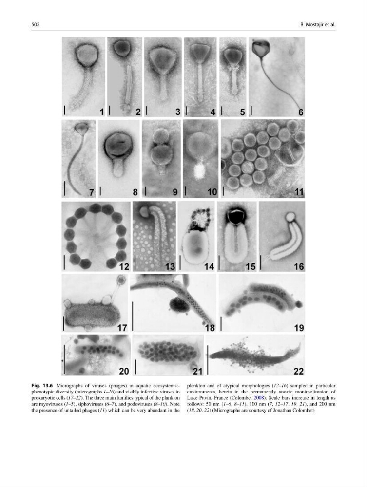
Fig. 13.6 Micrographs of viruses (phages) in aquatic ecosystems:phenotypic diversity (micrographs 1–16) and visibly infective viruses in
prokaryotic cells (17–22). The three main families typical of the plankton
are myoviruses (1–5), siphoviruses (6–7), and podoviruses (8–10). Note
the presence of untailed phages (11) which can be very abundant in the
plankton and of atypical morphologies (12–16) sampled in particular
environments, herein in the permanently anoxic monimolimnion of
Lake Pavin, France (Colombet 2008). Scale bars increase in length as
follows: 50 nm (1–6, 8–11), 100 nm (7, 12–17, 19, 21), and 200 nm
(18, 20, 22) (Micrographs are courtesy of Jonathan Colombet)
502
B. Mostajir et al.
prokaryotic cells (17–22). The three main families typical of the plankton
are myoviruses (1–5), siphoviruses (6–7), and podoviruses (8–10). Note
the presence of untailed phages (11) which can be very abundant in the
plankton and of atypical morphologies (12–16) sampled in particular
environments, herein in the permanently anoxic monimolimnion of
Lake Pavin, France (Colombet 2008). Scale bars increase in length as
follows: 50 nm (1–6, 8–11), 100 nm (7, 12–17, 19, 21), and 200 nm
(18, 20, 22) (Micrographs are courtesy of Jonathan Colombet)
502
B. Mostajir et al.