

cell. The activation of the basal body with membrane energy
from the proton gradient allows rotation flagellum that
drives the movement of the cell (Fig. 3.4). According to
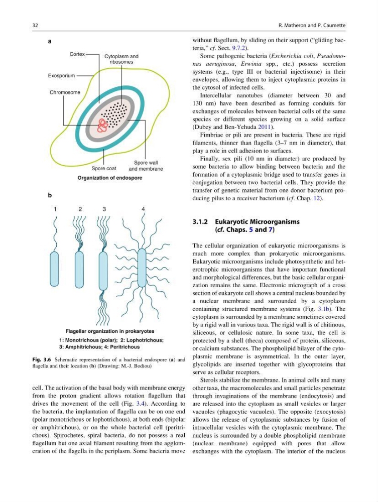
the bacteria, the implantation of flagella can be on one end
(polar monotrichous or lophotrichous), at both ends (bipolar
or amphitrichous), or on the whole bacterial cell (peritrichous). Spirochetes, spiral bacteria, do not possess a real
flagellum but one axial filament resulting from the agglomeration of the flagella in the periplasm. Some bacteria move
without flagellum, by sliding on their support (“gliding bacteria,” cf. Sect. 9.7.2).
Some pathogenic bacteria (Escherichia coli, Pseudomonas aeruginosa, Erwinia spp., etc.) possess secretion
systems (e.g., type III or bacterial injectisome) in their
envelopes, allowing them to inject cytoplasmic proteins in
the cytosol of infected cells.
Intercellular nanotubes (diameter between 30 and
130 nm) have been described as forming conduits for
exchanges of molecules between bacterial cells of the same
species or different species growing on a solid surface
(Dubey and Ben-Yehuda 2011).
Fimbriae or pili are present in bacteria. These are rigid
filaments, thinner than flagella (3–7 nm in diameter), that
play a role in cell adhesion to surfaces.
Finally, sex pili (10 nm in diameter) are produced by
some bacteria to allow binding between bacteria and the
formation of a cytoplasmic bridge used to transfer genes in
conjugation between two bacterial cells. They provide the
transfer of genetic material from one donor bacterium producing pilus to a receiver bacterium (cf. Chap. 12).
3.1.2 Eukaryotic Microorganisms
(cf. Chaps. 5 and 7)
The cellular organization of eukaryotic microorganisms is
much more complex than prokaryotic microorganisms.
Eukaryotic microorganisms include photosynthetic and heterotrophic microorganisms that have important functional
and morphological differences, but the basic cellular organization remains the same. Electronic micrograph of a cross
section of eukaryote cell shows a central nucleus bounded by
a nuclear membrane and surrounded by a cytoplasm
containing structured membrane systems (Fig. 3.1b). The
cytoplasm is surrounded by a membrane sometimes covered
by a rigid wall in various taxa. The rigid wall is of chitinous,
siliceous, or cellulosic nature. In some taxa, the cell is
protected by a shell (theca) composed of protein, siliceous,
or calcium substances. The phospholipid bilayer of the cytoplasmic membrane is asymmetrical. In the outer layer,
glycolipids are inserted together with glycoproteins that
serve as cellular receptors.
Sterols stabilize the membrane. In animal cells and many
other taxa, the macromolecules and small particles penetrate
through invaginations of the membrane (endocytosis) and
are released into the cytoplasm as small vesicles or larger
vacuoles (phagocytic vacuoles). The opposite (exocytosis)
allows the release of cytoplasmic substances by fusion of
intracellular vesicles with the cytoplasmic membrane. The
nucleus is surrounded by a double phospholipid membrane
(nuclear membrane) equipped with pores that allow
exchanges with the cytoplasm. The interior of the nucleus
Cytoplasm and
ribosomes
Chromosome
Cortex
Exosporium
Spore wall
and membrane
Spore coat
1
2
3
4
Organization of endospore
Flagellar organization in prokaryotes
1: Monotrichous (polar); 2: Lophotrichous;
3: Amphitrichous; 4: Peritrichous
a
b
Fig. 3.6 Schematic representation of a bacterial endospore (a) and
flagella and their location (b) (Drawing: M.-J. Bodiou)
32
R. Matheron and P. Caumette
from the proton gradient allows rotation flagellum that
drives the movement of the cell (Fig. 3.4). According to
the bacteria, the implantation of flagella can be on one end
(polar monotrichous or lophotrichous), at both ends (bipolar
or amphitrichous), or on the whole bacterial cell (peritrichous). Spirochetes, spiral bacteria, do not possess a real
flagellum but one axial filament resulting from the agglomeration of the flagella in the periplasm. Some bacteria move
without flagellum, by sliding on their support (“gliding bacteria,” cf. Sect. 9.7.2).
Some pathogenic bacteria (Escherichia coli, Pseudomonas aeruginosa, Erwinia spp., etc.) possess secretion
systems (e.g., type III or bacterial injectisome) in their
envelopes, allowing them to inject cytoplasmic proteins in
the cytosol of infected cells.
Intercellular nanotubes (diameter between 30 and
130 nm) have been described as forming conduits for
exchanges of molecules between bacterial cells of the same
species or different species growing on a solid surface
(Dubey and Ben-Yehuda 2011).
Fimbriae or pili are present in bacteria. These are rigid
filaments, thinner than flagella (3–7 nm in diameter), that
play a role in cell adhesion to surfaces.
Finally, sex pili (10 nm in diameter) are produced by
some bacteria to allow binding between bacteria and the
formation of a cytoplasmic bridge used to transfer genes in
conjugation between two bacterial cells. They provide the
transfer of genetic material from one donor bacterium producing pilus to a receiver bacterium (cf. Chap. 12).
3.1.2 Eukaryotic Microorganisms
(cf. Chaps. 5 and 7)
The cellular organization of eukaryotic microorganisms is
much more complex than prokaryotic microorganisms.
Eukaryotic microorganisms include photosynthetic and heterotrophic microorganisms that have important functional
and morphological differences, but the basic cellular organization remains the same. Electronic micrograph of a cross
section of eukaryote cell shows a central nucleus bounded by
a nuclear membrane and surrounded by a cytoplasm
containing structured membrane systems (Fig. 3.1b). The
cytoplasm is surrounded by a membrane sometimes covered
by a rigid wall in various taxa. The rigid wall is of chitinous,
siliceous, or cellulosic nature. In some taxa, the cell is
protected by a shell (theca) composed of protein, siliceous,
or calcium substances. The phospholipid bilayer of the cytoplasmic membrane is asymmetrical. In the outer layer,
glycolipids are inserted together with glycoproteins that
serve as cellular receptors.
Sterols stabilize the membrane. In animal cells and many
other taxa, the macromolecules and small particles penetrate
through invaginations of the membrane (endocytosis) and
are released into the cytoplasm as small vesicles or larger
vacuoles (phagocytic vacuoles). The opposite (exocytosis)
allows the release of cytoplasmic substances by fusion of
intracellular vesicles with the cytoplasmic membrane. The
nucleus is surrounded by a double phospholipid membrane
(nuclear membrane) equipped with pores that allow
exchanges with the cytoplasm. The interior of the nucleus
Cytoplasm and
ribosomes
Chromosome
Cortex
Exosporium
Spore wall
and membrane
Spore coat
1
2
3
4
Organization of endospore
Flagellar organization in prokaryotes
1: Monotrichous (polar); 2: Lophotrichous;
3: Amphitrichous; 4: Peritrichous
a
b
Fig. 3.6 Schematic representation of a bacterial endospore (a) and
flagella and their location (b) (Drawing: M.-J. Bodiou)
32
R. Matheron and P. Caumette