

2. Other transporters, secondary active transporters, use the
energy of a proton or concentration gradient. Some are
symport transporters for which penetration of the substance (sugars, amino acids) is generally associated with
a positive gradient of protons or Na
+
, the transfer of
protons or Na
+ resulting in the passage of the active
substance (Fig. 3.4). Other carriers are antiport
transporters in which the transport of the substance is
associated with a negative gradient (against gradient) of
Na
+ or proton or of another substance, for example, the
penetration of malate is associated with the output of
lactate during the malolactic fermentation in lactobacilli.
3. During active transport, the transported molecules can be
chemically modified. The best known mechanism is the
transport of sugars (glucose, fructose, mannose) involving
the phosphotransferase system which phosphorylates the
sugars at the level of carrier and releases the
phosphorylated sugar in the cytoplasm; for example, in
the case of glucose transport, the phosphate group of
glucose-6-phosphate originates from the hydrolysis of the
phosphate bond of phosphoenolpyruvate.
In phototrophic purple bacteria, the cytoplasmic membrane,
which is folded on itself, is at the origin of complex structures in
the form of tubules, lamellae, folds, or vesicles according to
bacterial groups (Fig. 3.27). These invaginations, observed also
in nitrifying bacteria, increase the surface-to-cell volume ratio
and thus promote exchange and energy activities.
Cell Wall
The cell wall allows the maintenance of cell shape; it is less
complex in Gram-positive than in Gram-negative bacteria.
In bacteria staining Gram positive, the wall is made as a
thick and rigid peptidoglycan or murein (Fig. 3.5b). Peptidoglycan which represents 90 % of weight of the wall is a
very large polymer formed by the sequence of two
derivatives of amino sugars, N-acetylglucosamine (AG)
and N-acetylmuramic acid (AM). Residues of AM are
substituted by tetrapeptides (TP). Polypeptide bridges (e.g.,
pentaglycine bridge) connect the tetrapeptides between them
and thus form a rigid entanglement (Fig. 3.5a). Teichoic
acids consisting of phosphoglycerol or ribitol phosphate
polymers, which are sometimes associated with sugars and
alanine, are also involved in wall rigidity.
In bacteria staining Gram negative, the wall structure is
more complex (Fig. 3.5b). The peptidoglycan is thinner and
represents only 10 % of the wall; there is no teichoic acid. On
the external side, the wall is limited by a membrane, the outer
membrane (Fig. 3.5c). The outer layer of this membrane
contains chains of lipopolysaccharides (LPS) which extend
outside the membrane forming side chains (antigen O). The
LPS is the endotoxin of Gram-negative bacteria. The outer
membrane is a diffusion barrier less effective than the cytoplasmic membrane. Proteins form channels (porins) for nonspecific passage of small molecules. The larger molecules are
transported specifically through the membrane. The space
between the outer membrane and cytoplasmic membrane is
called the periplasmic space or periplasm. It contains many
enzymatic proteins and may represent up to 40 % of bacterial
volume (Fig. 3.5b). Some Gram-negative bacteria secrete into
the environment outer membrane nanovesicles formed by the
bacterial outer membrane and containing components of the
periplasm and cytoplasm (enzymes and toxins).
In archaea, the cell wall has a different composition and is
more variable. Some archaea have walls with a single thick
layer formed of various polymers such as pseudomurein in
which N-acetylgalactosamine replaces N-acetylglucosamine.
Others have walls consisting of polysaccharides or heteropolysaccharides which are sulfated or non-sulfated. The wall of
other archaea is formed of a layer or double layer of protein or
glycoprotein subunits (Madigan et al. 2010).
Some bacteria and archaea are devoid of wall; in this case,
the cytoplasmic membrane is the unique cellular envelope.
The prokaryotic microorganisms are frequently
surrounded by a mucous layer called the glycocalyx. This
layer is composed of polysaccharides which are often
associated with polypeptides and that are secreted and
accumulated around the cell. The glycocalyx may be a thin
mucous layer called EPS (“exopolysaccharides” or
“exopolymeric substances”) or a thick layer more or less
rigid, called the capsule. These highly hydrated exopolymers
allow cells to agglomerate into biofilms (cf. Sect. 9.7.3), to
P
P
P
Membran proteins
Phopholipid bilayer
p
a
Alcohol
Phosphate
Glycerol
Fatty acid
Polar
head
Apolar
tail
b
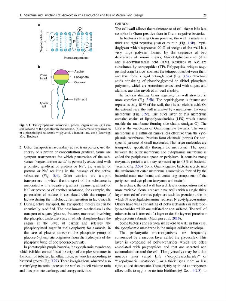
Fig. 3.3 The cytoplasmic membrane, general organization. (a) General scheme of the cytoplasmic membrane. (b) Schematic organization
of a phospholipid (alcohols ¼ glycerol, ethanolamine, etc.) (Drawing:
M.-J. Bodiou)
3 Structure and Functions of Microorganisms: Production and Use of Material and Energy
29
energy of a proton or concentration gradient. Some are
symport transporters for which penetration of the substance (sugars, amino acids) is generally associated with
a positive gradient of protons or Na
+
, the transfer of
protons or Na
+ resulting in the passage of the active
substance (Fig. 3.4). Other carriers are antiport
transporters in which the transport of the substance is
associated with a negative gradient (against gradient) of
Na
+ or proton or of another substance, for example, the
penetration of malate is associated with the output of
lactate during the malolactic fermentation in lactobacilli.
3. During active transport, the transported molecules can be
chemically modified. The best known mechanism is the
transport of sugars (glucose, fructose, mannose) involving
the phosphotransferase system which phosphorylates the
sugars at the level of carrier and releases the
phosphorylated sugar in the cytoplasm; for example, in
the case of glucose transport, the phosphate group of
glucose-6-phosphate originates from the hydrolysis of the
phosphate bond of phosphoenolpyruvate.
In phototrophic purple bacteria, the cytoplasmic membrane,
which is folded on itself, is at the origin of complex structures in
the form of tubules, lamellae, folds, or vesicles according to
bacterial groups (Fig. 3.27). These invaginations, observed also
in nitrifying bacteria, increase the surface-to-cell volume ratio
and thus promote exchange and energy activities.
Cell Wall
The cell wall allows the maintenance of cell shape; it is less
complex in Gram-positive than in Gram-negative bacteria.
In bacteria staining Gram positive, the wall is made as a
thick and rigid peptidoglycan or murein (Fig. 3.5b). Peptidoglycan which represents 90 % of weight of the wall is a
very large polymer formed by the sequence of two
derivatives of amino sugars, N-acetylglucosamine (AG)
and N-acetylmuramic acid (AM). Residues of AM are
substituted by tetrapeptides (TP). Polypeptide bridges (e.g.,
pentaglycine bridge) connect the tetrapeptides between them
and thus form a rigid entanglement (Fig. 3.5a). Teichoic
acids consisting of phosphoglycerol or ribitol phosphate
polymers, which are sometimes associated with sugars and
alanine, are also involved in wall rigidity.
In bacteria staining Gram negative, the wall structure is
more complex (Fig. 3.5b). The peptidoglycan is thinner and
represents only 10 % of the wall; there is no teichoic acid. On
the external side, the wall is limited by a membrane, the outer
membrane (Fig. 3.5c). The outer layer of this membrane
contains chains of lipopolysaccharides (LPS) which extend
outside the membrane forming side chains (antigen O). The
LPS is the endotoxin of Gram-negative bacteria. The outer
membrane is a diffusion barrier less effective than the cytoplasmic membrane. Proteins form channels (porins) for nonspecific passage of small molecules. The larger molecules are
transported specifically through the membrane. The space
between the outer membrane and cytoplasmic membrane is
called the periplasmic space or periplasm. It contains many
enzymatic proteins and may represent up to 40 % of bacterial
volume (Fig. 3.5b). Some Gram-negative bacteria secrete into
the environment outer membrane nanovesicles formed by the
bacterial outer membrane and containing components of the
periplasm and cytoplasm (enzymes and toxins).
In archaea, the cell wall has a different composition and is
more variable. Some archaea have walls with a single thick
layer formed of various polymers such as pseudomurein in
which N-acetylgalactosamine replaces N-acetylglucosamine.
Others have walls consisting of polysaccharides or heteropolysaccharides which are sulfated or non-sulfated. The wall of
other archaea is formed of a layer or double layer of protein or
glycoprotein subunits (Madigan et al. 2010).
Some bacteria and archaea are devoid of wall; in this case,
the cytoplasmic membrane is the unique cellular envelope.
The prokaryotic microorganisms are frequently
surrounded by a mucous layer called the glycocalyx. This
layer is composed of polysaccharides which are often
associated with polypeptides and that are secreted and
accumulated around the cell. The glycocalyx may be a thin
mucous layer called EPS (“exopolysaccharides” or
“exopolymeric substances”) or a thick layer more or less
rigid, called the capsule. These highly hydrated exopolymers
allow cells to agglomerate into biofilms (cf. Sect. 9.7.3), to
P
P
P
Membran proteins
Phopholipid bilayer
p
a
Alcohol
Phosphate
Glycerol
Fatty acid
Polar
head
Apolar
tail
b
Fig. 3.3 The cytoplasmic membrane, general organization. (a) General scheme of the cytoplasmic membrane. (b) Schematic organization
of a phospholipid (alcohols ¼ glycerol, ethanolamine, etc.) (Drawing:
M.-J. Bodiou)
3 Structure and Functions of Microorganisms: Production and Use of Material and Energy
29