

of about 1 millisecond during which the bacteria tumble on
itself before going in another direction; the displacement
due to the activation of flagellum is controlled by the
chemotaxis signaling systems (cf. Sect. 9.2.2). In liquid
medium, the speed of movement by the flagella varies
depending on cells: Bacillus megaterium (large rod)
moves from 1.5 to 2 mm/min, whereas Vibrio cholerae
(curved rod) can move at the speed of 12 mm/min which
is approximately 3,000 times the length of the bacterial
body. The generally accepted average is 2–4 mm/min.
• Bacteria possessing gas vacuoles
One adaptation to aquatic life is the synthesis of
flotation vesicle, through a set of proteins called “gas
vesicles proteins.” These vesicles are small organelles
found in different aquatic bacteria such as cyanobacteria
(Nostoc muscorum, Anabaena flos-aquae, etc.), heterotrophic bacteria [(Prosthecomicrobium pneumaticum,
Aquabacter, Ancylobacter aquaticus (Konopka et al.
1977)], phototrophic purple and green bacteria
(Thiocapsa, Thiodictyon, Pelodictyon, etc.), Firmicutes
(Bacillus megaterium), and also archaea (Halobacterium halobium, Haloferax sp.) (Halladay et al. 1992)
or even soil actinobacteria (van Keulen et al. 2005),
probably so that they can remain at the top of the water
column. The assembly of vesicles in the cell leads to the
formation of gas vacuoles visible under an optical
microscope as clear spaces in the cell. They provide
buoyancy by decreasing cell density. The vesicles that
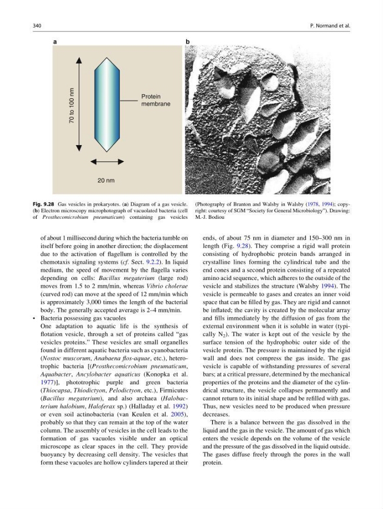
form these vacuoles are hollow cylinders tapered at their
ends, of about 75 nm in diameter and 150–300 nm in
length (Fig. 9.28). They comprise a rigid wall protein
consisting of hydrophobic protein bands arranged in
crystalline lines forming the cylindrical tube and the
end cones and a second protein consisting of a repeated
amino acid sequence, which adheres to the outside of the
vesicle and stabilizes the structure (Walsby 1994). The
vesicle is permeable to gases and creates an inner void
space that can be filled by gas. They are rigid and cannot
be inflated; the cavity is created by the molecular array
and fills immediately by the diffusion of gas from the
external environment when it is soluble in water (typically N 2 ). The water is kept out of the vesicle by the
surface tension of the hydrophobic outer side of the
vesicle protein. The pressure is maintained by the rigid
wall and does not compress the gas inside. The gas
vesicle is capable of withstanding pressures of several
bars; at a critical pressure, determined by the mechanical
properties of the proteins and the diameter of the cylindrical structure, the vesicle collapses permanently and
cannot return to its initial shape and be refilled with gas.
Thus, new vesicles need to be produced when pressure
decreases.
There is a balance between the gas dissolved in the
liquid and the gas in the vesicle. The amount of gas which
enters the vesicle depends on the volume of the vesicle
and the pressure of the gas dissolved in the liquid outside.
The gases diffuse freely through the pores in the wall
protein.
70 to 100 nm
20 nm
Protein
membrane
a
b
Fig. 9.28 Gas vesicles in prokaryotes. (a) Diagram of a gas vesicle.
(b) Electron microscopy microphotograph of vacuolated bacteria (cell
of Prosthecomicrobium pneumaticum) containing gas vesicles
(Photography of Branton and Walsby in Walsby (1978, 1994); copyright: courtesy of SGM “Society for General Microbiology”). Drawing:
M.-J. Bodiou
340
P. Normand et al.
itself before going in another direction; the displacement
due to the activation of flagellum is controlled by the
chemotaxis signaling systems (cf. Sect. 9.2.2). In liquid
medium, the speed of movement by the flagella varies
depending on cells: Bacillus megaterium (large rod)
moves from 1.5 to 2 mm/min, whereas Vibrio cholerae
(curved rod) can move at the speed of 12 mm/min which
is approximately 3,000 times the length of the bacterial
body. The generally accepted average is 2–4 mm/min.
• Bacteria possessing gas vacuoles
One adaptation to aquatic life is the synthesis of
flotation vesicle, through a set of proteins called “gas
vesicles proteins.” These vesicles are small organelles
found in different aquatic bacteria such as cyanobacteria
(Nostoc muscorum, Anabaena flos-aquae, etc.), heterotrophic bacteria [(Prosthecomicrobium pneumaticum,
Aquabacter, Ancylobacter aquaticus (Konopka et al.
1977)], phototrophic purple and green bacteria
(Thiocapsa, Thiodictyon, Pelodictyon, etc.), Firmicutes
(Bacillus megaterium), and also archaea (Halobacterium halobium, Haloferax sp.) (Halladay et al. 1992)
or even soil actinobacteria (van Keulen et al. 2005),
probably so that they can remain at the top of the water
column. The assembly of vesicles in the cell leads to the
formation of gas vacuoles visible under an optical
microscope as clear spaces in the cell. They provide
buoyancy by decreasing cell density. The vesicles that
form these vacuoles are hollow cylinders tapered at their
ends, of about 75 nm in diameter and 150–300 nm in
length (Fig. 9.28). They comprise a rigid wall protein
consisting of hydrophobic protein bands arranged in
crystalline lines forming the cylindrical tube and the
end cones and a second protein consisting of a repeated
amino acid sequence, which adheres to the outside of the
vesicle and stabilizes the structure (Walsby 1994). The
vesicle is permeable to gases and creates an inner void
space that can be filled by gas. They are rigid and cannot
be inflated; the cavity is created by the molecular array
and fills immediately by the diffusion of gas from the
external environment when it is soluble in water (typically N 2 ). The water is kept out of the vesicle by the
surface tension of the hydrophobic outer side of the
vesicle protein. The pressure is maintained by the rigid
wall and does not compress the gas inside. The gas
vesicle is capable of withstanding pressures of several
bars; at a critical pressure, determined by the mechanical
properties of the proteins and the diameter of the cylindrical structure, the vesicle collapses permanently and
cannot return to its initial shape and be refilled with gas.
Thus, new vesicles need to be produced when pressure
decreases.
There is a balance between the gas dissolved in the
liquid and the gas in the vesicle. The amount of gas which
enters the vesicle depends on the volume of the vesicle
and the pressure of the gas dissolved in the liquid outside.
The gases diffuse freely through the pores in the wall
protein.
70 to 100 nm
20 nm
Protein
membrane
a
b
Fig. 9.28 Gas vesicles in prokaryotes. (a) Diagram of a gas vesicle.
(b) Electron microscopy microphotograph of vacuolated bacteria (cell
of Prosthecomicrobium pneumaticum) containing gas vesicles
(Photography of Branton and Walsby in Walsby (1978, 1994); copyright: courtesy of SGM “Society for General Microbiology”). Drawing:
M.-J. Bodiou
340
P. Normand et al.