

41
Streptococcaceae Representatives
Lactococcus garvieae (= Enterococcus seriolicida)
Characteristics of the Disease
Infection of ayu with this pathogen may be exacerbated by prior infection/infestation with other organisms, such as blood fl ukes, that may weaken the host (Kumon
et al. 2002 ). Internal signs of disease were absent in golden shiners, although raised
lesions were apparent on the body surface (Robinson and Meyer 1966 ). Yellowtails
were damaged in the liver, kidney, spleen and intestine, and there was a concomitant
accumulation of ascitic fl uid in the peritoneal cavity (Kusuda et al. 1976 ; 1991 ;
Ugajin 1981 ). In rainbow trout, the disease was of sudden onset, and was described
as a hyperacute systemic disease (Eldar and Ghittino 1999 ); haemorrhaging in the
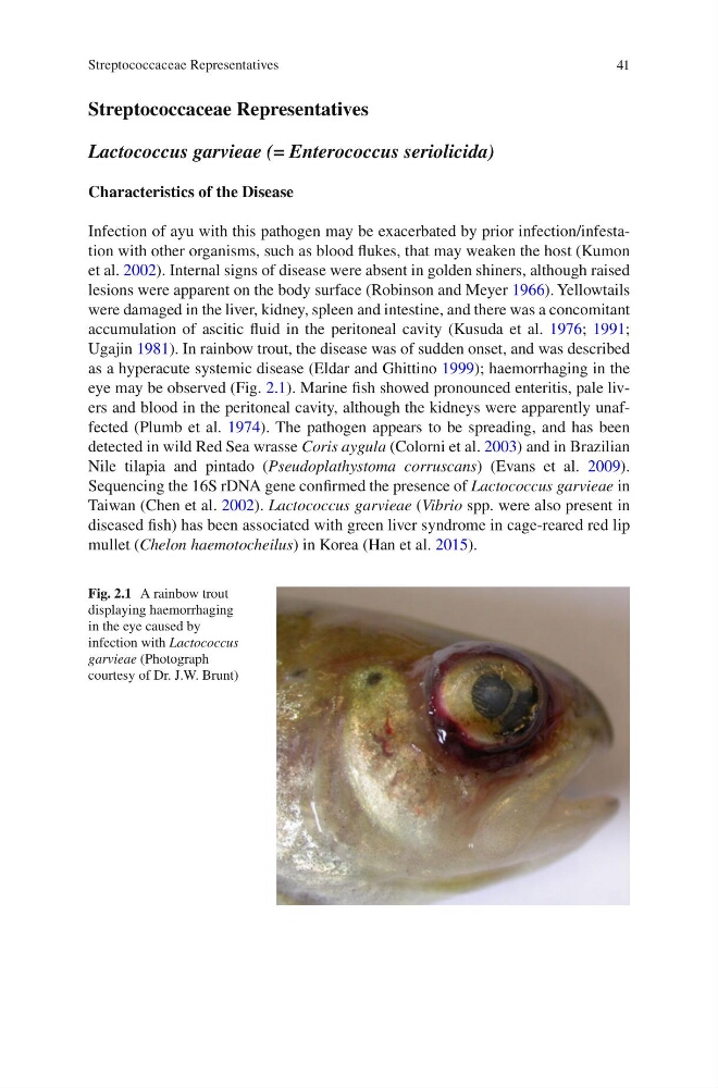
eye may be observed (Fig. 2.1 ). Marine fi sh showed pronounced enteritis, pale livers and blood in the peritoneal cavity, although the kidneys were apparently unaffected (Plumb et al. 1974 ). The pathogen appears to be spreading, and has been
detected in wild Red Sea wrasse Coris aygula (Colorni et al. 2003 ) and in Brazilian
Nile tilapia and pintado ( Pseudoplathystoma corruscans ) (Evans et al. 2009 ).
Sequencing the 16S rDNA gene confi rmed the presence of Lactococcus garvieae in
Taiwan (Chen et al. 2002 ). Lactococcus garvieae ( Vibrio spp. were also present in
diseased fi sh) has been associated with green liver syndrome in cage-reared red lip
mullet ( Chelon haemotocheilus ) in Korea (Han et al. 2015 ).
Fig. 2.1 A rainbow trout
displaying haemorrhaging
in the eye caused by
infection with Lactococcus
garvieae (Photograph
courtesy of Dr. J.W. Brunt)
Streptococcaceae Representatives
Streptococcaceae Representatives
Lactococcus garvieae (= Enterococcus seriolicida)
Characteristics of the Disease
Infection of ayu with this pathogen may be exacerbated by prior infection/infestation with other organisms, such as blood fl ukes, that may weaken the host (Kumon
et al. 2002 ). Internal signs of disease were absent in golden shiners, although raised
lesions were apparent on the body surface (Robinson and Meyer 1966 ). Yellowtails
were damaged in the liver, kidney, spleen and intestine, and there was a concomitant
accumulation of ascitic fl uid in the peritoneal cavity (Kusuda et al. 1976 ; 1991 ;
Ugajin 1981 ). In rainbow trout, the disease was of sudden onset, and was described
as a hyperacute systemic disease (Eldar and Ghittino 1999 ); haemorrhaging in the
eye may be observed (Fig. 2.1 ). Marine fi sh showed pronounced enteritis, pale livers and blood in the peritoneal cavity, although the kidneys were apparently unaffected (Plumb et al. 1974 ). The pathogen appears to be spreading, and has been
detected in wild Red Sea wrasse Coris aygula (Colorni et al. 2003 ) and in Brazilian
Nile tilapia and pintado ( Pseudoplathystoma corruscans ) (Evans et al. 2009 ).
Sequencing the 16S rDNA gene confi rmed the presence of Lactococcus garvieae in
Taiwan (Chen et al. 2002 ). Lactococcus garvieae ( Vibrio spp. were also present in
diseased fi sh) has been associated with green liver syndrome in cage-reared red lip
mullet ( Chelon haemotocheilus ) in Korea (Han et al. 2015 ).
Fig. 2.1 A rainbow trout
displaying haemorrhaging
in the eye caused by
infection with Lactococcus
garvieae (Photograph
courtesy of Dr. J.W. Brunt)
Streptococcaceae Representatives