

713
ulceration in cyprinids, after which motile aeromonads exacerbate and may lead to
the development of a systemic disease during which time the initial pathogen has
effectively disappeared. Certainly, co-infection which may be attributed to more
than one pathogen is likely to have been underreported. Nevertheless, there is evidence for the presence of two or more “pathogens” within some diseased fi sh (Loch
et al. 2012 ). Diseased Chinook salmon ( Oncorhynchus tshawytscha ), which were
returning to spawn in tributaries associated with Lake Michigan, USA, were found
to be populated by multiple bacterial pathogens (Loch et al. 2012 ). Specifi c
examples:
• Uncultured obligately intracellular chlamydia-like organisms associated with
epitheliocystis may be associated with co-infections. Specifi cally, ‘ Candidatus
Piscichlamydia salmonis’ and ‘ Candidatus Clavochlamydia salmonicola’ have
been attributed with co-infections both in the same population and within the
same individuals of wild brown trout in Switzerland (Schmidt-Posthaus et al.
2012 ).
• Sometimes, a second organism may be a secondary invader, such as the case with
Aer. hydrophila , which was found in fi sh from lakes in Saskatchewan, Canada
with columnaris (Scott and Bollinger 2014 ).
• Possibly, the presence of one pathogen/parasite renders the host more susceptible
to another as was considered in the case of parasitism by Ichthyophthirius multifi liis leading to greater susceptibility to and invasion of hybrid tilapia with Fla.
columnare (Xu et al. 2014 ).
• Dual infection of Ren. salmoninarum and Nanophyetus salminicola Chapin in
wild juvenile Chinook salmon has been recorded (Sandell et al. 2015 ).
• Neoparamoeba perurans , which causes amoebic gill disease, and Y. ruckeri have
been indicated to co-infect Atlantic salmon in Tasmania, Australia (ValdenegroVega et al. 2015 ).
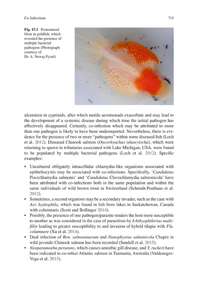
Fig. 15.1 Pronounced
bloat in goldfi sh, which
revealed the presence of
multiple bacterial
pathogens (Photograph
courtesy of
Dr. A. Newaj-Fyzul)
Co-Infections
ulceration in cyprinids, after which motile aeromonads exacerbate and may lead to
the development of a systemic disease during which time the initial pathogen has
effectively disappeared. Certainly, co-infection which may be attributed to more
than one pathogen is likely to have been underreported. Nevertheless, there is evidence for the presence of two or more “pathogens” within some diseased fi sh (Loch
et al. 2012 ). Diseased Chinook salmon ( Oncorhynchus tshawytscha ), which were
returning to spawn in tributaries associated with Lake Michigan, USA, were found
to be populated by multiple bacterial pathogens (Loch et al. 2012 ). Specifi c
examples:
• Uncultured obligately intracellular chlamydia-like organisms associated with
epitheliocystis may be associated with co-infections. Specifi cally, ‘ Candidatus
Piscichlamydia salmonis’ and ‘ Candidatus Clavochlamydia salmonicola’ have
been attributed with co-infections both in the same population and within the
same individuals of wild brown trout in Switzerland (Schmidt-Posthaus et al.
2012 ).
• Sometimes, a second organism may be a secondary invader, such as the case with
Aer. hydrophila , which was found in fi sh from lakes in Saskatchewan, Canada
with columnaris (Scott and Bollinger 2014 ).
• Possibly, the presence of one pathogen/parasite renders the host more susceptible
to another as was considered in the case of parasitism by Ichthyophthirius multifi liis leading to greater susceptibility to and invasion of hybrid tilapia with Fla.
columnare (Xu et al. 2014 ).
• Dual infection of Ren. salmoninarum and Nanophyetus salminicola Chapin in
wild juvenile Chinook salmon has been recorded (Sandell et al. 2015 ).
• Neoparamoeba perurans , which causes amoebic gill disease, and Y. ruckeri have
been indicated to co-infect Atlantic salmon in Tasmania, Australia (ValdenegroVega et al. 2015 ).
Fig. 15.1 Pronounced
bloat in goldfi sh, which
revealed the presence of
multiple bacterial
pathogens (Photograph
courtesy of
Dr. A. Newaj-Fyzul)
Co-Infections