

666
opaqueness; Hyperaemic stomach; Haemorrhaging in/on the internal organs;
Brain damage; Blood in the cranium; Emaciation; Pale, elongated/swollen
spleen; Pale (possibly mottled/discoloured) liver; Yellowish liver (with hyperaemic areas); Swollen liver; Generalised liquefaction; The presence of
tumours
Histopathological Examination of Diseased Tissues
Although many histological procedures may be routinely used, it is important for
the bacteriologist that Gram-stained sections should be prepared. Thus, the presence
of any offending bacterial pathogen will quickly be recognised. Microscopic examination of Gram-stained material will enable the determination of the basic staining
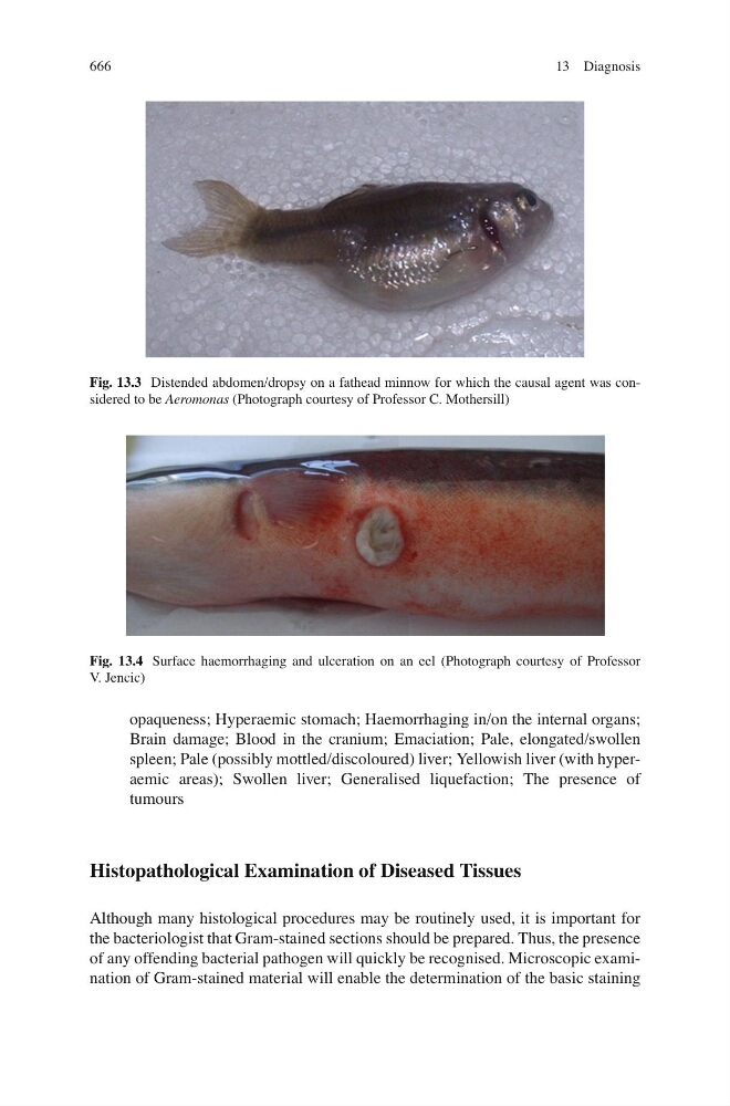
Fig. 13.4 Surface haemorrhaging and ulceration on an eel (Photograph courtesy of Professor
V. Jencic)
Fig. 13.3 Distended abdomen/dropsy on a fathead minnow for which the causal agent was considered to be Aeromonas (Photograph courtesy of Professor C. Mothersill)
13 Diagnosis