

218
it is most frequently located on the fl anks. Infected fi sh exhibit inappetance, and
appear darker in colour. Secondary invasion of the ulcer by fungi or other bacteria
is common. If the fi sh recovers, the healed ulcer is recognisable as a grey-black scar.
Frequently, contraction of the collagen of the scar tissue can result in serious deformity, which reduces the commercial value of the fi sh (Fijan 1972 ). In some instances,
CE may also result in generalised septicaemia and death. Unlike furunculosis,
which usually occurs only when water temperatures exceed 16 °C, CE may occur at
all water temperatures.
Aer. salmonicida subsp. masoucida has been associated with extensive haemorrhagic septicaemia, including the presence of surface and muscle haemorrhaging, in
black rockfi sh ( Sebastes schlegelii ) from South Korea (Fig. 5.4 ).
Aer. salmonicida has been reported to cause a cutaneous ulcerative disease in
ornamental fi sh (Figs. 5.5 , 5.6 ), especially goldfi sh ( Carassius auratus ), where the
condition is referred at goldfi sh ulcer disease, which is a stress-mediated condition
associated with the atypical form of the pathogen (Dror et al. 2006 ). However, the
disease has been known for a long time previously. Ulcer disease of cyprinids, in
general, has occurred in widely separated geographical locations, including the
USA, Japan and England (Shotts et al. 1980 ). Mawdesley-Thomas ( 1969 ) studied,
in detail, an outbreak of an ulcerative disease of goldfi sh, and recovered Aer. salmonicida . Symptoms included lethargy, loss of orientation, and abnormal swimming behaviour. The ulcers were of various sizes and depths, and some fi sh died
shortly after infection. Secondary invasion of the ulcers by Saprolegnia was
observed. More recently, we have recovered an extremely fastidious form of the
pathogen from ulcerated carp, goldfi sh and roach in England. Here, there was
evidence of secondary invasion of the ulcers by Aer. hydrophila . According to
McCarthy and Roberts ( 1980 ), ulcer disease differed from CE in the following
ways:
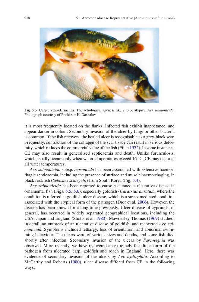
Fig. 5.3 Carp erythrodermatitis. The aetiological agent is likely to be atypical Aer. salmonicida.
Photograph courtesy of Professor H. Daskalov
5 Aeromonadaceae Representative (Aeromonas salmonicida)
it is most frequently located on the fl anks. Infected fi sh exhibit inappetance, and
appear darker in colour. Secondary invasion of the ulcer by fungi or other bacteria
is common. If the fi sh recovers, the healed ulcer is recognisable as a grey-black scar.
Frequently, contraction of the collagen of the scar tissue can result in serious deformity, which reduces the commercial value of the fi sh (Fijan 1972 ). In some instances,
CE may also result in generalised septicaemia and death. Unlike furunculosis,
which usually occurs only when water temperatures exceed 16 °C, CE may occur at
all water temperatures.
Aer. salmonicida subsp. masoucida has been associated with extensive haemorrhagic septicaemia, including the presence of surface and muscle haemorrhaging, in
black rockfi sh ( Sebastes schlegelii ) from South Korea (Fig. 5.4 ).
Aer. salmonicida has been reported to cause a cutaneous ulcerative disease in
ornamental fi sh (Figs. 5.5 , 5.6 ), especially goldfi sh ( Carassius auratus ), where the
condition is referred at goldfi sh ulcer disease, which is a stress-mediated condition
associated with the atypical form of the pathogen (Dror et al. 2006 ). However, the
disease has been known for a long time previously. Ulcer disease of cyprinids, in
general, has occurred in widely separated geographical locations, including the
USA, Japan and England (Shotts et al. 1980 ). Mawdesley-Thomas ( 1969 ) studied,
in detail, an outbreak of an ulcerative disease of goldfi sh, and recovered Aer. salmonicida . Symptoms included lethargy, loss of orientation, and abnormal swimming behaviour. The ulcers were of various sizes and depths, and some fi sh died
shortly after infection. Secondary invasion of the ulcers by Saprolegnia was
observed. More recently, we have recovered an extremely fastidious form of the
pathogen from ulcerated carp, goldfi sh and roach in England. Here, there was
evidence of secondary invasion of the ulcers by Aer. hydrophila . According to
McCarthy and Roberts ( 1980 ), ulcer disease differed from CE in the following
ways:
Fig. 5.3 Carp erythrodermatitis. The aetiological agent is likely to be atypical Aer. salmonicida.
Photograph courtesy of Professor H. Daskalov
5 Aeromonadaceae Representative (Aeromonas salmonicida)