

162
F. J. R. TAYLOR:
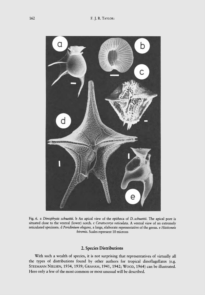
Fig. 6. a Dinophysis schuettii. b An apical view of the epitheca of D. schuettii. The apical pore is
situated close to the ventral (lower) notch. c Ceratocorys reticuiata. A ventral view of an extremely
reticulated specimen. d Peridinium eiegans, a large, elaborate representative of the genus. e Histioneis
biremis. Scales represent 10 microns
2. Species Distributions
With such a wealth of species, it is not surprising that representatives of virtually all
the types of distributions found by other authors for tropical dinoflagellates (e.g.
STEEMANN NIELSEN, 1934, 1939; GRAHAM, 1941, 1942; WOOD, 1964) can be illustrated.
Here only a few of the most common or most unusual will be described.
F. J. R. TAYLOR:
Fig. 6. a Dinophysis schuettii. b An apical view of the epitheca of D. schuettii. The apical pore is
situated close to the ventral (lower) notch. c Ceratocorys reticuiata. A ventral view of an extremely
reticulated specimen. d Peridinium eiegans, a large, elaborate representative of the genus. e Histioneis
biremis. Scales represent 10 microns
2. Species Distributions
With such a wealth of species, it is not surprising that representatives of virtually all
the types of distributions found by other authors for tropical dinoflagellates (e.g.
STEEMANN NIELSEN, 1934, 1939; GRAHAM, 1941, 1942; WOOD, 1964) can be illustrated.
Here only a few of the most common or most unusual will be described.