

74
for rigidity of their cartilaginous skeletons. The neural or hemal arches and centra
of vertebrae, as well as the chondrocranium, jaws and visceral arches, in the
supporting cartilages of fi ns and clasper spurs, are possible locations where tesserae
in sharks may occur (Applegate 1967 ). The structure of shark’s tesserae with respect
to their outer and inner surfaces is not homogenous. It was observed by Kemp and
Westrin ( 1979 ) using SEM that “calcospherites and hydroxyapatite crystals similar
to those commonly seen on the surface of bone are present on the outer surface of
the tessera adjacent to the perichondrium. On the inner surface adjoining hyaline
cartilage, however, calcospherites of variable size are the predominant surface feature. TEM shows calcifi cation in close association with coarse collagen fi brils on
the outer side of a tessera, but such fi brils are absent from the cartilaginous matrix
along the underside of tesserae,” (Kemp and Westrin 1979 ).
The tesserate patterns in modern Chondrichthyes are also an example of the
primitive characteristic. Calcifi cation phenomenon observed in this fi sh group
“may be derived from an ancestral pattern of a continuous bed of calcifi ed cartilage underlying a layer of perichondral bone as theorized by Ørvig ( 1951 ),”
(Kemp and Westrin 1979 ).
The structure and ultrastructure of calcifi ed cartilage in the endoskeletal tesserae have
been characterized not only of sharks (Bargmann 1939 ; Kemp and Westrin 1979 ; Dean
et al. 2008 ) but also of stingrays (Summers et al. 1998 ; Dean and Summers 2006 ; Dean
2007 ; Dean et al. 2007 , 2009a , b , 2010 ). For example, growth of the jaw cartilage as well
as the tessellation of the round stingray Urobatis halleri has been recently studied in the
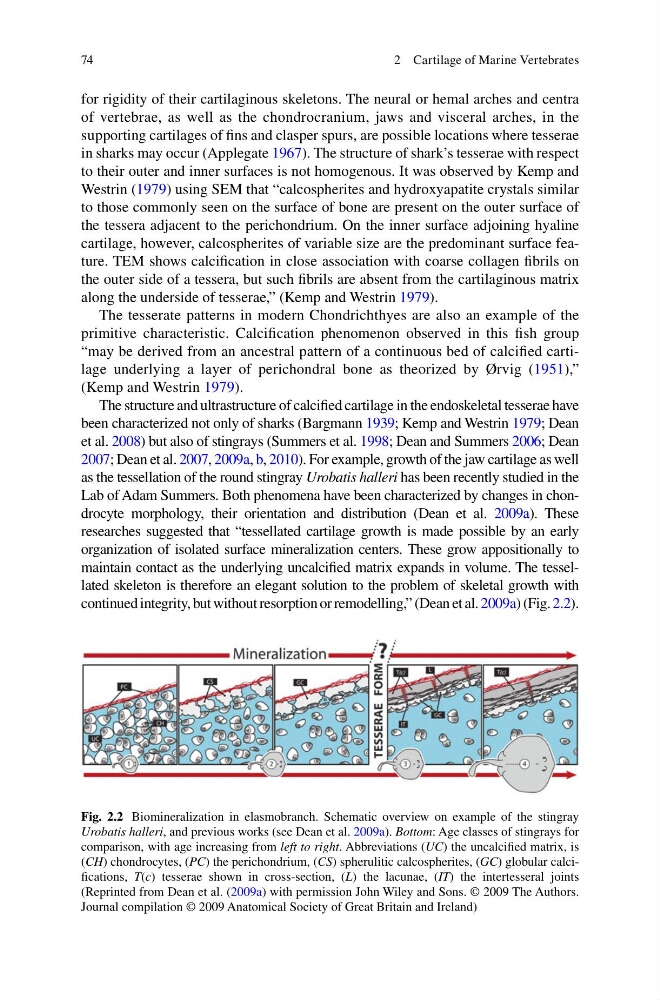
Lab of Adam Summers. Both phenomena have been characterized by changes in chondrocyte morphology, their orientation and distribution (Dean et al. 2009a ). These
researches suggested that “tessellated cartilage growth is made possible by an early
organization of isolated surface mineralization centers. These grow appositionally to
maintain contact as the underlying uncalcifi ed matrix expands in volume. The tessellated skeleton is therefore an elegant solution to the problem of skeletal growth with
continued integrity, but without resorption or remodelling,” (Dean et al. 2009a ) (Fig. 2.2 ).
Fig. 2.2 Biomineralization in elasmobranch. Schematic overview on example of the stingray
Urobatis halleri , and previous works (see Dean et al. 2009a ). Bottom : Age classes of stingrays for
comparison, with age increasing from left to right . Abbreviations ( UC ) the uncalcifi ed matrix, is
( CH ) chondrocytes, ( PC ) the perichondrium, ( CS ) spherulitic calcospherites, ( GC ) globular calcifi cations, T ( c ) tesserae shown in cross-section, ( L ) the lacunae, ( IT ) the intertesseral joints
(Reprinted from Dean et al. ( 2009a ) with permission John Wiley and Sons. © 2009 The Authors.
Journal compilation © 2009 Anatomical Society of Great Britain and Ireland)
2 Cartilage of Marine Vertebrates
for rigidity of their cartilaginous skeletons. The neural or hemal arches and centra
of vertebrae, as well as the chondrocranium, jaws and visceral arches, in the
supporting cartilages of fi ns and clasper spurs, are possible locations where tesserae
in sharks may occur (Applegate 1967 ). The structure of shark’s tesserae with respect
to their outer and inner surfaces is not homogenous. It was observed by Kemp and
Westrin ( 1979 ) using SEM that “calcospherites and hydroxyapatite crystals similar
to those commonly seen on the surface of bone are present on the outer surface of
the tessera adjacent to the perichondrium. On the inner surface adjoining hyaline
cartilage, however, calcospherites of variable size are the predominant surface feature. TEM shows calcifi cation in close association with coarse collagen fi brils on
the outer side of a tessera, but such fi brils are absent from the cartilaginous matrix
along the underside of tesserae,” (Kemp and Westrin 1979 ).
The tesserate patterns in modern Chondrichthyes are also an example of the
primitive characteristic. Calcifi cation phenomenon observed in this fi sh group
“may be derived from an ancestral pattern of a continuous bed of calcifi ed cartilage underlying a layer of perichondral bone as theorized by Ørvig ( 1951 ),”
(Kemp and Westrin 1979 ).
The structure and ultrastructure of calcifi ed cartilage in the endoskeletal tesserae have
been characterized not only of sharks (Bargmann 1939 ; Kemp and Westrin 1979 ; Dean
et al. 2008 ) but also of stingrays (Summers et al. 1998 ; Dean and Summers 2006 ; Dean
2007 ; Dean et al. 2007 , 2009a , b , 2010 ). For example, growth of the jaw cartilage as well
as the tessellation of the round stingray Urobatis halleri has been recently studied in the
Lab of Adam Summers. Both phenomena have been characterized by changes in chondrocyte morphology, their orientation and distribution (Dean et al. 2009a ). These
researches suggested that “tessellated cartilage growth is made possible by an early
organization of isolated surface mineralization centers. These grow appositionally to
maintain contact as the underlying uncalcifi ed matrix expands in volume. The tessellated skeleton is therefore an elegant solution to the problem of skeletal growth with
continued integrity, but without resorption or remodelling,” (Dean et al. 2009a ) (Fig. 2.2 ).
Fig. 2.2 Biomineralization in elasmobranch. Schematic overview on example of the stingray
Urobatis halleri , and previous works (see Dean et al. 2009a ). Bottom : Age classes of stingrays for
comparison, with age increasing from left to right . Abbreviations ( UC ) the uncalcifi ed matrix, is
( CH ) chondrocytes, ( PC ) the perichondrium, ( CS ) spherulitic calcospherites, ( GC ) globular calcifi cations, T ( c ) tesserae shown in cross-section, ( L ) the lacunae, ( IT ) the intertesseral joints
(Reprinted from Dean et al. ( 2009a ) with permission John Wiley and Sons. © 2009 The Authors.
Journal compilation © 2009 Anatomical Society of Great Britain and Ireland)
2 Cartilage of Marine Vertebrates