

72
manner that they do not touch other cells. Cell–matrix interactions in the mammalian
cartilage are essential for the maintenance of the extracellular matrix. The organic
network that contains fi brillar collagen provides the capacity to contain the swelling
pressure of the embedded proteoglycans as well as tensile strength to the tissue.
Cartilaginous collagen fi brils are represented by collagens type II, IX and XI. Typical
representatives of the proteoglycans are biglycan, fi bromodulin, lumican and epiphycan, the cell surface syndecans and glypican, the basement membrane proteoglycan, perlecan, and the small leucine-rich proteoglycans decorin. Both decorin
and type IX collagen play important role in regulation of collagen fi bril formation.
One of the large chondroitin sulphate-containing proteoglycans is the aggrecan that
is expressed during chondrogenesis. The aggrecan is responsible for achievement of
osmotic resistance necessary for cartilage to resist compressive loads. “Following
its secretion, aggrecan self-assembles into a supramolecular structure with as many
as 50 monomers bound to a fi lament of hyaluronan” (Knudson and Knudson 2001 ).
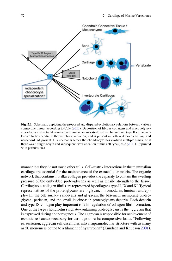
Fig. 2.1 Schematic depicting the proposed and disputed evolutionary relations between various
connective tissues according to Cole ( 2011 ). Deposition of fi brous collagens and mucopolysaccharides in a structured connective tissue is an ancestral feature. In contrast, type II collagen is
known to be specifi c to the vertebrate radiation, and is present in both vertebrate cartilage and
notochord. At present it is unclear whether the chondrocyte has evolved multiple times, or if
there was a single origin and subsequent diversifi cation of this cell type (Cole ( 2011 ). Reprinted
with permission.)
2 Cartilage of Marine Vertebrates
manner that they do not touch other cells. Cell–matrix interactions in the mammalian
cartilage are essential for the maintenance of the extracellular matrix. The organic
network that contains fi brillar collagen provides the capacity to contain the swelling
pressure of the embedded proteoglycans as well as tensile strength to the tissue.
Cartilaginous collagen fi brils are represented by collagens type II, IX and XI. Typical
representatives of the proteoglycans are biglycan, fi bromodulin, lumican and epiphycan, the cell surface syndecans and glypican, the basement membrane proteoglycan, perlecan, and the small leucine-rich proteoglycans decorin. Both decorin
and type IX collagen play important role in regulation of collagen fi bril formation.
One of the large chondroitin sulphate-containing proteoglycans is the aggrecan that
is expressed during chondrogenesis. The aggrecan is responsible for achievement of
osmotic resistance necessary for cartilage to resist compressive loads. “Following
its secretion, aggrecan self-assembles into a supramolecular structure with as many
as 50 monomers bound to a fi lament of hyaluronan” (Knudson and Knudson 2001 ).
Fig. 2.1 Schematic depicting the proposed and disputed evolutionary relations between various
connective tissues according to Cole ( 2011 ). Deposition of fi brous collagens and mucopolysaccharides in a structured connective tissue is an ancestral feature. In contrast, type II collagen is
known to be specifi c to the vertebrate radiation, and is present in both vertebrate cartilage and
notochord. At present it is unclear whether the chondrocyte has evolved multiple times, or if
there was a single origin and subsequent diversifi cation of this cell type (Cole ( 2011 ). Reprinted
with permission.)
2 Cartilage of Marine Vertebrates