

380
major part is involved in synthesis of spine-like structures termed “pharyngeal
teeth”. Stratum corneum, stratum germinativum and stratum spinosum are three
layers of the epithelium. The scaly keratin of possibly sloughing cells is the main
component of the spine’s surface. The more than 100 μm thick cornifi ed layer is
characteristic for the spine and can reach three to seven times the depth of corresponding layers in other parts. As natural alternative to marine turtle without toothed
jaws, pharyngeal teeth are probably used for breaking food into small fragments
(Yoshie and Honma 1976 ).
Comparative study on alpha- and beta- keratins in reptiles was initiated by
Alexander ( 1970 ). From functional point of view, “α-keratins are crucial for water
conservation (stratum corneum) in amniotes and for heat conservation (hair and fur)
in mammals. Therefore these versatile biological materials likely played a crucial
role in the successful invasion of terrestrial habitats by vertebrates,” (Fudge et al.
2009b ). Toni et al. ( 2007 ) characterized both types of these structural proteins as
follow:
“The epidermis of reptiles […] contains relatively large α-keratins of 40–68 kDa,
and beta-keratins of smaller molecular weight (10–20 kDa) that possess different
chemical and physical properties. α-keratins or cytokeratins constitute the
intermediate- fi lament bundles and are present in most epidermal layers where they
have a role in mechanical resistance, impart form to cells, or determine their changes
in shape. Alpha-keratins are contained in keratinocytes of the alpha-layer of the
epidermis, which mainly functions in permitting epidermal stretching and as a barrier against water-loss. Differently from alpha-keratins, beta-keratins are specifi c
proteins utilized to make hard structures including scales, scutes, and claws (beak
and feathers in birds). Beta-keratins are produced in the pre-corneous layer of the
specialized epidermis of these skin appendages. The importance of beta-keratins in
reptilian scales and their relatively small dimensions has led to recent studies of
these proteins in detail; and some of their genes identifi ed, sequenced, and cloned,”
(Toni et al. 2007 ).
Two types of keratinocyte differentiation has been described in the epidermis of
turtles. So called softer keratinization was observed in the limbs, neck and tail, and
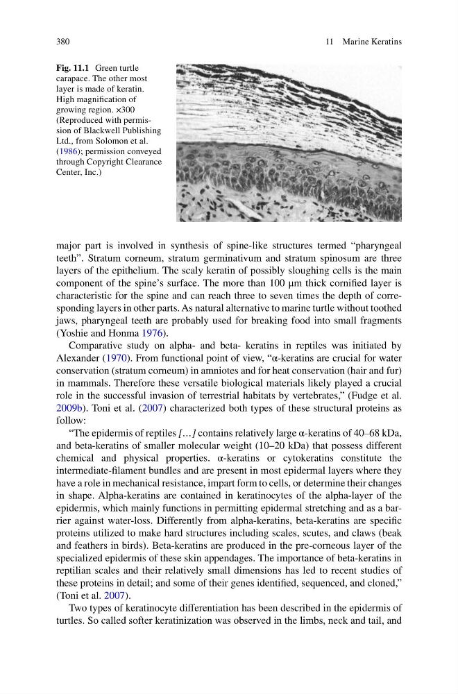
Fig. 11.1 Green turtle
carapace. The other most
layer is made of keratin.
High magnifi cation of
growing region. ×300
(Reproduced with permission of Blackwell Publishing
Ltd., from Solomon et al.
( 1986 ); permission conveyed
through Copyright Clearance
Center, Inc.)
11 Marine Keratins
major part is involved in synthesis of spine-like structures termed “pharyngeal
teeth”. Stratum corneum, stratum germinativum and stratum spinosum are three
layers of the epithelium. The scaly keratin of possibly sloughing cells is the main
component of the spine’s surface. The more than 100 μm thick cornifi ed layer is
characteristic for the spine and can reach three to seven times the depth of corresponding layers in other parts. As natural alternative to marine turtle without toothed
jaws, pharyngeal teeth are probably used for breaking food into small fragments
(Yoshie and Honma 1976 ).
Comparative study on alpha- and beta- keratins in reptiles was initiated by
Alexander ( 1970 ). From functional point of view, “α-keratins are crucial for water
conservation (stratum corneum) in amniotes and for heat conservation (hair and fur)
in mammals. Therefore these versatile biological materials likely played a crucial
role in the successful invasion of terrestrial habitats by vertebrates,” (Fudge et al.
2009b ). Toni et al. ( 2007 ) characterized both types of these structural proteins as
follow:
“The epidermis of reptiles […] contains relatively large α-keratins of 40–68 kDa,
and beta-keratins of smaller molecular weight (10–20 kDa) that possess different
chemical and physical properties. α-keratins or cytokeratins constitute the
intermediate- fi lament bundles and are present in most epidermal layers where they
have a role in mechanical resistance, impart form to cells, or determine their changes
in shape. Alpha-keratins are contained in keratinocytes of the alpha-layer of the
epidermis, which mainly functions in permitting epidermal stretching and as a barrier against water-loss. Differently from alpha-keratins, beta-keratins are specifi c
proteins utilized to make hard structures including scales, scutes, and claws (beak
and feathers in birds). Beta-keratins are produced in the pre-corneous layer of the
specialized epidermis of these skin appendages. The importance of beta-keratins in
reptilian scales and their relatively small dimensions has led to recent studies of
these proteins in detail; and some of their genes identifi ed, sequenced, and cloned,”
(Toni et al. 2007 ).
Two types of keratinocyte differentiation has been described in the epidermis of
turtles. So called softer keratinization was observed in the limbs, neck and tail, and
Fig. 11.1 Green turtle
carapace. The other most
layer is made of keratin.
High magnifi cation of
growing region. ×300
(Reproduced with permission of Blackwell Publishing
Ltd., from Solomon et al.
( 1986 ); permission conveyed
through Copyright Clearance
Center, Inc.)
11 Marine Keratins