

367
Arterial System and Heart The pericardial cavity is the space where the bulbus
arteriosus is located in teleost. The question concerning the arterial or cardiac
nature of this organ is still open (Benjamin et al. 1983 ). The description of bulbus
arteriosus is to be found in the work by Braun et al. ( 2003 ):
“Typically, it is swollen proximally, tapering distally into the ventral aorta.
Within these parameters, a large variety of shapes occur, but the functional signifi cance
of the different shapes is unknown. The internal morphology of the bulbus also
shows a large amount of variance between species. The lumen can be smooth,
ridged or trabeculated. The trabeculae can be “spongy”, or separated into discrete
longitudinal and radial elements. The bulbar wall is composed of three layers: an
intima composed of a single layer of endothelial cells, a thick media primarily
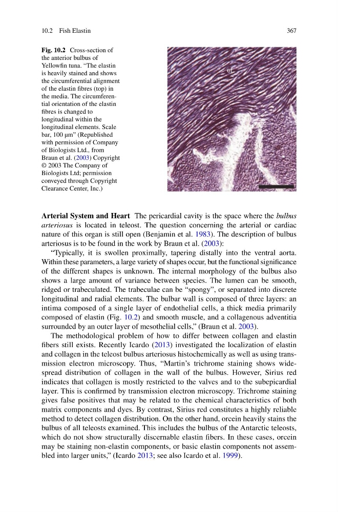
composed of elastin (Fig. 10.2 ) and smooth muscle, and a collagenous adventitia
surrounded by an outer layer of mesothelial cells,” (Braun et al. 2003 ).
The methodological problem of how to differ between collagen and elastin
fi bers still exists. Recently Icardo ( 2013 ) investigated the localization of elastin
and collagen in the teleost bulbus arteriosus histochemically as well as using transmission electron microscopy. Thus, “Martin’s trichrome staining shows widespread distribution of collagen in the wall of the bulbus. However, Sirius red
indicates that collagen is mostly restricted to the valves and to the subepicardial
layer. This is confi rmed by transmission electron microscopy. Trichrome staining
gives false positives that may be related to the chemical characteristics of both
matrix components and dyes. By contrast, Sirius red constitutes a highly reliable
method to detect collagen distribution. On the other hand, orcein heavily stains the
bulbus of all teleosts examined. This includes the bulbus of the Antarctic teleosts,
which do not show structurally discernable elastin fi bers. In these cases, orcein
may be staining non- elastin components, or basic elastin components not assembled into larger units,” (Icardo 2013 ; see also Icardo et al. 1999 ).
Fig. 10.2 Cross-section of
the anterior bulbus of
Yellowfi n tuna. “The elastin
is heavily stained and shows
the circumferential alignment
of the elastin fi bres (top) in
the media. The circumferential orientation of the elastin
fi bres is changed to
longitudinal within the
longitudinal elements. Scale
bar, 100 µm” (Republished
with permission of Company
of Biologists Ltd., from
Braun et al. ( 2003 ) Copyright
© 2003 The Company of
Biologists Ltd; permission
conveyed through Copyright
Clearance Center, Inc.)
10.2 Fish Elastin
Arterial System and Heart The pericardial cavity is the space where the bulbus
arteriosus is located in teleost. The question concerning the arterial or cardiac
nature of this organ is still open (Benjamin et al. 1983 ). The description of bulbus
arteriosus is to be found in the work by Braun et al. ( 2003 ):
“Typically, it is swollen proximally, tapering distally into the ventral aorta.
Within these parameters, a large variety of shapes occur, but the functional signifi cance
of the different shapes is unknown. The internal morphology of the bulbus also
shows a large amount of variance between species. The lumen can be smooth,
ridged or trabeculated. The trabeculae can be “spongy”, or separated into discrete
longitudinal and radial elements. The bulbar wall is composed of three layers: an
intima composed of a single layer of endothelial cells, a thick media primarily
composed of elastin (Fig. 10.2 ) and smooth muscle, and a collagenous adventitia
surrounded by an outer layer of mesothelial cells,” (Braun et al. 2003 ).
The methodological problem of how to differ between collagen and elastin
fi bers still exists. Recently Icardo ( 2013 ) investigated the localization of elastin
and collagen in the teleost bulbus arteriosus histochemically as well as using transmission electron microscopy. Thus, “Martin’s trichrome staining shows widespread distribution of collagen in the wall of the bulbus. However, Sirius red
indicates that collagen is mostly restricted to the valves and to the subepicardial
layer. This is confi rmed by transmission electron microscopy. Trichrome staining
gives false positives that may be related to the chemical characteristics of both
matrix components and dyes. By contrast, Sirius red constitutes a highly reliable
method to detect collagen distribution. On the other hand, orcein heavily stains the
bulbus of all teleosts examined. This includes the bulbus of the Antarctic teleosts,
which do not show structurally discernable elastin fi bers. In these cases, orcein
may be staining non- elastin components, or basic elastin components not assembled into larger units,” (Icardo 2013 ; see also Icardo et al. 1999 ).
Fig. 10.2 Cross-section of
the anterior bulbus of
Yellowfi n tuna. “The elastin
is heavily stained and shows
the circumferential alignment
of the elastin fi bres (top) in
the media. The circumferential orientation of the elastin
fi bres is changed to
longitudinal within the
longitudinal elements. Scale
bar, 100 µm” (Republished
with permission of Company
of Biologists Ltd., from
Braun et al. ( 2003 ) Copyright
© 2003 The Company of
Biologists Ltd; permission
conveyed through Copyright
Clearance Center, Inc.)
10.2 Fish Elastin