

362
related to physico-chemical (Mistrali et al. 1971 ; Gosline 1978 ; Lillie and Gosline
1996 ; Lillie et al. 1996 ) and mechanical (Gosline and French 1979 ; Aaron and
Gosline 1981 ; Lillie and Gosline 1990 ; Lillie et al. 1994 ) properties, as well as the
structural biology (Montes 1996 ) and genomics (He et al. 2007 ) of this bioelastomer. Special attention was paid to biomechanics of arterial elastin (see for details
Gundiah et al. 2007 , 2009 ). Tropoelastin is a 60–72 kDa protein of the extracellular
matrix origin that is distributed in all vertebrates, with exception of cyclostomes
(Wise and Weiss 2009 ). Molecular orientation of tropoelastin is determined by surface hydrophobicity (Le Brun et al. 2012 ).
Wise and Weiss ( 2009 ) characterized some properties of this protein as follow:
“Secreted tropoelastin is tethered to the cell surface, where it aggregates into
organised spheres for cross-linking and incorporation into growing elastic fi bres.
Tropoelastin is characterised by alternating hydrophobic and hydrophilic domains,
and is highly fl exible. The conserved C-terminus is an area of the molecule of particular biological importance in that it is required for both incorporation into elastin,
and for cellular interactions. Mature cross-linked tropoelastin gives elastin, which
confers resilience and elasticity on a diverse range of tissues,” (Wise and Weiss
2009 ; see also Sandberg et al. 1971 ).
According to the modern view (see for review Yeo et al. 2012 ), tropoelastin is
secreted by elastogenic cells such as smooth muscle cells, endothelial cells, and
fi broblasts. At the cell surface, the monomers cluster through hydrophobic domain
interactions in an aqueous environment by the entropically driven process of coacervation. These tropoelastin assemblies remain attached through the C terminus to
cell-surface integrin αvβ3 and glycosaminoglycans until deposition on microfi brillar scaffolds, which direct the shape and orientation of elastic fi bers. Microfi brillar
proteins recruit lysyl oxidase, which reacts with specifi c tropoelastin lysine residues
to form cross-links. These cross-links occur at multiple sites in the molecule and are
enriched in domains 19–25. Cross-linking imposes expansional constraints on elastin and renders elastic fi bers resilient under repetitive mechanical stretching (Yeo
et al. 2012 ).
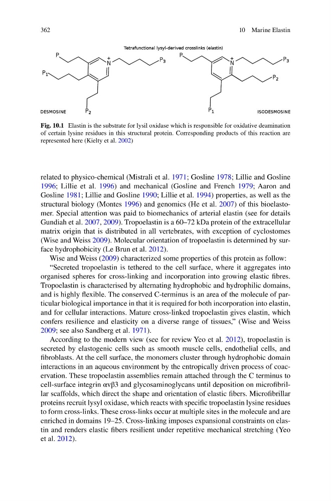
Fig. 10.1 Elastin is the substrate for lysil oxidase which is responsible for oxidative deamination
of certain lysine residues in this structural protein. Corresponding products of this reaction are
represented here (Kielty et al. 2002 )
10 Marine Elastin
related to physico-chemical (Mistrali et al. 1971 ; Gosline 1978 ; Lillie and Gosline
1996 ; Lillie et al. 1996 ) and mechanical (Gosline and French 1979 ; Aaron and
Gosline 1981 ; Lillie and Gosline 1990 ; Lillie et al. 1994 ) properties, as well as the
structural biology (Montes 1996 ) and genomics (He et al. 2007 ) of this bioelastomer. Special attention was paid to biomechanics of arterial elastin (see for details
Gundiah et al. 2007 , 2009 ). Tropoelastin is a 60–72 kDa protein of the extracellular
matrix origin that is distributed in all vertebrates, with exception of cyclostomes
(Wise and Weiss 2009 ). Molecular orientation of tropoelastin is determined by surface hydrophobicity (Le Brun et al. 2012 ).
Wise and Weiss ( 2009 ) characterized some properties of this protein as follow:
“Secreted tropoelastin is tethered to the cell surface, where it aggregates into
organised spheres for cross-linking and incorporation into growing elastic fi bres.
Tropoelastin is characterised by alternating hydrophobic and hydrophilic domains,
and is highly fl exible. The conserved C-terminus is an area of the molecule of particular biological importance in that it is required for both incorporation into elastin,
and for cellular interactions. Mature cross-linked tropoelastin gives elastin, which
confers resilience and elasticity on a diverse range of tissues,” (Wise and Weiss
2009 ; see also Sandberg et al. 1971 ).
According to the modern view (see for review Yeo et al. 2012 ), tropoelastin is
secreted by elastogenic cells such as smooth muscle cells, endothelial cells, and
fi broblasts. At the cell surface, the monomers cluster through hydrophobic domain
interactions in an aqueous environment by the entropically driven process of coacervation. These tropoelastin assemblies remain attached through the C terminus to
cell-surface integrin αvβ3 and glycosaminoglycans until deposition on microfi brillar scaffolds, which direct the shape and orientation of elastic fi bers. Microfi brillar
proteins recruit lysyl oxidase, which reacts with specifi c tropoelastin lysine residues
to form cross-links. These cross-links occur at multiple sites in the molecule and are
enriched in domains 19–25. Cross-linking imposes expansional constraints on elastin and renders elastic fi bers resilient under repetitive mechanical stretching (Yeo
et al. 2012 ).
Fig. 10.1 Elastin is the substrate for lysil oxidase which is responsible for oxidative deamination
of certain lysine residues in this structural protein. Corresponding products of this reaction are
represented here (Kielty et al. 2002 )
10 Marine Elastin